>Link back: HIGHLIGHTS, Quantum Materials, Symmetry & its Applications, Ferroelectrics, Piezoelectrics & Multiferroics, Semiconductor Fibers & Metalattices.
Ultrafast and Nonlinear Optics
________________________________
Optical creation of a supercrystal with three-dimensional nanoscale periodicity, V. A. Stoica, N. Laanait, C. Dai, Z. Hong, Z. Zhang, S. Lei, M. R. McCarter, A. Yadav, A. R. Damodaran, S. Das, G. A. Stone, J. Karapetrova, D. A. Walko, X. Zhang, L. W. Martin, R. Ramesh, L-Q. Chen, H. Wen, V. Gopalan, J. W. Freeland, Nature Materials, 18, 377 (2019).
Stimulation with ultrafast light pulses is a means to realize and manipulate new states of matter. While non-equilibrium optical excitations can lead to transient phases with emergent structural, electronic, and magnetic phenomena, a significant challenge is to stabilize them as persistent states. Here, we show that atomic scale PbTiO3/SrTiO3 superlattices that delicately counterpoise strain and polarization states in alternate layers, undergo a light-stimulated transition to a new polar supercrystal phase, which is triggered by sub-picosecond excitation with above bandgap energy light. The resulting supercrystal persists indefinitely under ambient conditions, has not been created via equilibrium routes, and can be erased by heating above a critical temperature. Using X-ray scattering and microscopy, we reveal that this unusual and complex phase consists of long-range, coherent three-dimensional (3-D) structure with nanoscale periodicities of up to 30 nm, consisting of polar, strain, and charge cooperative ordering. Phase-field model describes this emergent phase as a photo-induced charge-stabilized supercrystal that is triggered from a mixed two-phase thermal equilibrium state. Our results demonstrate new opportunities for light activated pathways to thermally inaccessible and emergent metastable states.
 Supercrystal observation by X-ray diffraction and microscopy: a. (left and middle) Diffraction along the K-L (qy-qz) plane for the mixed phase (FE + V) pristine sample shows evidence of order only along the z direction with distinct peaks due to the FE and V phases, as noted by the horizontal lines. b. Upon optical excitation with light above the PTO bandgap, a periodic 2-dimensional diffraction pattern appears due to spatial ordering in the y-z plane. (right) A 3-D view of a representative PTO layer where in-plane strain (εy) distribution is overlaid with polar displacement vector maps. Along the x-y plane, the polar vortex order alternates between in-plane and out-of-plane orientation for their respective vortex axes.
__________
Linear and nonlinear optical probe of the ferroelectric-like phase transition in a polar metal LiOsO3, Haricharan Padmanabhan, Yoonsang Park, Danilo Puggioni, Yakun Yuan, Yanwei Cao, Lev Gasparov, Youguo Shi, Jak Chakhalian, James M. Rondinelli, and Venkatraman Gopalan, Phys. Review Lett. 113, 122906 (2018).
LiOsO3 is one of the first materials identified in the recent literature as a “polar metal,” a class of materials that are simultaneously noncentrosymmetric and metallic. In this work, the linear and nonlinear optical susceptibility of LiOsO3 is studied by means of ellipsometry and optical second harmonic generation (SHG). Strong optical birefringence is observed using spectroscopic ellipsometry. The nonlinear optical susceptibility extracted from SHG polarimetry reveals that the tensor components are of the same magnitude as in the isostructural insulator LiNbO3, except the component along the polar axis d33 is suppressed by an order of magnitude. Temperature-dependent SHG measurements in combination with Raman spectroscopy indicate a continuous order-disorder type polar phase transition at 140 K. Linear and nonlinear optical microscopy measurements reveal 109 deg /71 deg ferroelastic domain walls, like in other trigonal ferroelectrics. No 180 deg polar domain walls are observed to emerge across the phase transition.
 Ferroelastic domains and phase transition in polar metal, LiOsO3 with (a) showing a linear optical micrograph at 300 K and (b) a nonlinear optical micrograph using SHG reflectance at 20 K. A schematic of the domains is shown below, with the arrows denoting the direction of the [100] crystallographic axis, obtained from electron back-scattering diffraction. The scale bar in (a) is 10 μm. (c) The temperature dependence of optical second harmonic generation, SHG, is shown in the upper panel, with an enlarged plot of the dependence near the phase transition shown in the inset. The lower panel shows the Raman linewidths of three different phonon modes as a function of temperature, with 2E, 5E, and 7E corresponding to modes at frequencies of 206 cm-1, 402 cm-1, and 492 cm-1, respectively, at room temperature.
__________
Light-activated Gigahertz ferroelectric domain dynamics, Hirofumi Akamatsu, Yakun Yuan, Vladimir A. Stoica, Greg Stone, Tiannan Yang, Zijian Hong, Shiming Lei, Yi Zhu, Ryan C. Haislmaier, John W. Freeland, Long-Qing Chen, Haidan Wen, and Venkatraman Gopalan, Physical Review Letters120, 096101 (2018).
Using time- and spatially resolved hard x-ray diffraction microscopy, the striking structural and electrical dynamics upon optical excitation of a single crystal of BaTiO3 are simultaneously captured on subnanoseconds and nanoscale within individual ferroelectric domains and across walls. A large emergent photoinduced electric field of up to 20×10^6 V/m is discovered in a surface layer of the crystal, which then drives polarization and lattice dynamics that are dramatically distinct in a surface layer versus bulk regions. A dynamical phase-field modeling method is developed that reveals the microscopic origin of these dynamics, leading to gigahertz polarization and elastic waves traveling in the crystal with sonic speeds and spatially varying frequencies. The advances in spatio-temporal imaging and dynamical modeling tools open up opportunities for disentangling ultrafast processes in complex mesoscale structures such as ferroelectric domains.
 A spatially resolved pump-probe experiment and domain configuration of BaTiO3 single crystal sample. (a) (Inset) The incident plane. The tilting angles of domains χ and φ are also shown. a and c domains are depicted in green (polarization in the crystal plane depicted by double-headed black arrows) and orange (polarization perpendicular to the surface), respectively. (b) The experimental and theoretical vertical strain, Δε33, as a function of time, in the surface and subsurface domains.
__________
Emergent Low‐Symmetry Phases and Large Property Enhancements in Ferroelectric KNbO3 Bulk Crystals, Tom TA Lummen, J Leung, Amit Kumar, X Wu, Y Ren, Brian K VanLeeuwen, Ryan C Haislmaier, Martin Holt, Keji Lai, Sergei V Kalinin, Venkatraman Gopalan, Advanced Materials, 29, (2017).
The design of new or enhanced functionality in materials is traditionally viewed as requiring the discovery of new chemical compositions through synthesis. Large property enhancements may however also be hidden within already well-known materials, when their structural symmetry is deviated from equilibrium through a small local strain or field. This work reports on the discovery of enhanced material properties associated with a new metastable phase of monoclinic symmetry within bulk KNbO3. This phase is found to co-exist with the nominal orthorhombic phase at room temperature, and is both induced by and stabilized with local strains generated by a network of ferroelectric domain walls. While the local microstructural shear strain involved is only ~0.017%, the concurrent symmetry reduction results in an optical second harmonic generation response that is over 550% higher at room temperature. Moreover, the meandering walls of the low symmetry domains also exhibit enhanced electrical conductivity on the order of 1 S m-1. This discovery reveals a potential new route to local engineering of significant property enhancements and conductivity through symmetry lowering in ferroelectric crystals.
 Emergent monoclinic phase in KNbO3 crystals with large property enhancements: Scanning SHG microscopy image ( ) of a densely twinned domain structure in a KNbO3 single crystal (surface ∥ (001)pc). The image is composed of several stitched area scans (10% overlap). Scale bar: 50 µm. The proposed polarization directions in the various domains are depicted in the zoom-in insets (see text). The zero-signal area on the bottom left corresponds to a thin Ag marker pad that serves as a positional reference (see Experimental Section).
__________
Ultrafast quasiparticle dynamics in correlated semimetal Ca3Ru2O7, Yakun Yuan, Peter Kissin, Danilo Puggioni, Kevin Cremin, Shiming Lei, Yu Wang, Zhiqiang Mao, James M Rondinelli, Richard D Averitt, Venkatraman Gopalan, arXiv preprint arXiv:1901.02512, Physical Review B, 99, 155111(2019).
The correlated polar semimetal Ca3Ru2O7 exhibits a rich phase diagram including two magnetic transitions (TN = 56 K and TC = 48 K) with the appearance of an insulating-like pseudogap (at TC ). In addition, there is a crossover back to metallic behavior at T* = 30 K, the origin of which is still under debate. We utilized ultrafast optical-pump optical-probe spectroscopy to investigate quasiparticle dynamics as a function of temperature in this enigmatic quantum material. We identify two dynamical processes, both of which are influenced by the onset of the pseudogap. This includes electron-phonon relaxation and, below TC, the onset of a phonon bottleneck hindering the relaxation of quasiparticles across the pseudogap. We introduce a gap-modified two-temperature model to describe the temperature dependence of electron-phonon thermalization, and use the Rothwarf-Taylor to model the phonon bottleneck. In conjunction with density functional theory, our experimental results synergistically reveal the origin of the T -dependent pseudogap. Further, our data and analysis indicate that T* emerges as a natural consequence of T -dependent gapping out of carriers, and does not correspond to a separate electronic transition. Our results highlight the value of low-fluence ultrafast optics as a sensitive probe of low-energy electronic structure, thermodynamic parameters, and transport properties of Ruddlesden-Popper ruthenates.
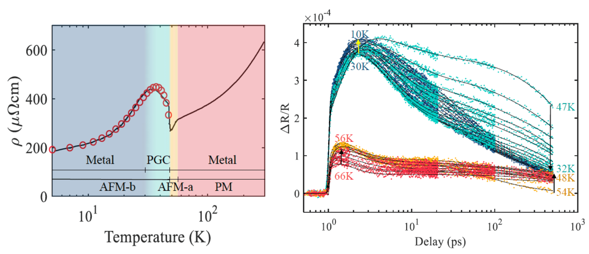 Ultrafast dynamics in Ca3Ru2O7: (Left) dc resistivity of Ca3Ru2O7 in the a-b plane. Regions with different colors represent different behaviors emerging at low temperature. PM stands for paramagnetic phase, AFM for antiferromagnetic phases, and PGC for pseudogap region. The red line is a fit based on T -dependent carrier concentration and scattering rate. (Right) Temperature-dependent optical pump-optical probe data on Ca3Ru2O7 . Four distinct relaxation processes are clearly observed at various temperatures, color-coded to match the resistivity phase diagram in (b). Black lines are fits using the multiexponential decay model described in the paper.
_________
>Link back: HIGHLIGHTS, Quantum Materials, Symmetry & its Applications, Ferroelectrics, Piezoelectrics & Multiferroics, Semiconductor Fibers & Metalattices.