Category Archives: Sample preparation questions

Detergent removal 1: Gel-assisted proteolysis

As promised, here’s a straightforward way to remove detergents, urea, and other LC-MS incompatible nasties from small-volume samples. The literature calls it ‘gel-assisted’ proteolysis. The idea is to entrap the protein solution in a polyacrylamide gel matrix, wash out detergents, salts, and chaotropic agents, and perform in-gel digestion. This technique works great for membrane proteins which are notoriously difficult to dissolve, and it is quite useful for any protein sample clean-up.

Slide1

For my little demo, I used a 1 mg/mL BSA solution in 2% SDS. The disulfides were reduced with TCEP and alkylated with IAA, after which the protein solution was very thoroughly mixed with a 30% T acrylamide monomer solution. I quickly added 10% APS and TEMED and immediately vortexed and centrifuged this mixture so that the liquid is collected at the bottom of the tube. The polymerization time is very short, a minute or two! I left it to completely polymerize for another 20 min.

Slide2

Using scalpel I removed the gel plug from the tube and diced it into small pieces. After 6 washes with 8 mg/mL ammonium bicarbonate in 50% acetonitrile, I dried the gel pieces in neat acetonitrile, removed the acetonitrile and added trypsin (see the in-gel digestion tutorial for details).


Fast forward to the MS analysis: Since the original BSA solution was very concentrated, I dissolved the peptides in 540 uL of mobile phase and injected 1 uL of this solution (55 ng total protein on column or approximately 1 pmol).

Slide3


Second example shows an ion chromatogram from a 10-uL IP eluent containing 2% detergent which I cleaned up and digested using this technique.

Slide4


Slide5


Slide6


As always, let me know if you have questions!

References:

doi: 10.1074/mcp.M500138-MCP200

doi: 10.1074/mcp.M800068-MCP200


LC-MS compatible detergents

They do exist! They are compatible with the reverse-phase chromatography! They solubilize stubborn proteins and improve proteolysis! Here are their names:

Acid-labile surfactants are hydrolyzed at low pH, and the hydrolysis products are compatible with reversed-phase separations and MS. These include RapiGest SF and PPS Silent Surfactant.

Invitrosol is a homogeneous surfactant whose elution profile does not overlap with the proteolytic peptides elution profiles.

ProteaseMAX is a surfactant that degrades during proteolysis, and its degradation products do not interfere with LC-MS.

All other detergents are not compatible with LC-MS and must be removed from the samples prior to analysis. These detergents include SDS (sodium dodecyl sulfate) and LDS (Lithium dodecyl sulfate), NP-40, Triton, Octyl glucoside and octyl thioglucoside, CHAPS, sodium deoxycholate, lauryl maltoside, Brij-35, etc. There are several ways to remove detergents, which is a topic for another blog post!

Extracting large peptides from gel after digestion

First, let’s define ‘large’. The Proteome Discoverer sets the upper limit for a precursor ion at 10,000 Da. This means anything bigger than 10 kDa will not be considered even if it’s present in the MS data, and a different software package ($$) will be required to analyze the high MW data. Clearly, proteolytic peptides with MW > 10 kDa will not be very useful for protein identification. I suggest using a different enzyme or a combination of enzymes. I have seen tryptic peptides up to 7 kDa in some in-gel digested samples, so apparently some large peptides do come out of gel.

Next, a large peptide’s physico-chemical properties (e.g. hydrophobicity, pI, hydrodynamic radius) must be considered as they will affect the extraction efficiency. If the peptide’s properties are known, the extraction solvent composition and pH can be adjusted to improve the peptide’s solubility.

Finally, let’s consider the gel from which the large peptides need to come out. Obviously, it will be easier to get the large peptides out of a 4 %T gel than out of a 20 %T one. Soaking a gel piece in deionized water and then freezing it should crash enough pores in the gel to improve the extraction  of large peptides (water expands as it freezes). Additionally, the gel could be ‘squeezed out’ a few times by changing extraction solvent from neat acetonitrile to an aqueous mixture. The gel piece will shrink in acetonitrile expelling the peptide solution. Re-hydrating the gel and then shrinking it again in acetonitrile will ‘squeeze out’ more of the digest.

Using elevated temperature (50 C), vortex mixer, and/or ultrasonic bath should all improve the extraction. Use common sense: 50C and a high pH buffer is not a good idea for the phosphopeptide extraction. Another word of caution: don’t get carried away. Three extraction steps should be enough. If you end up with a large volume (e.g. more than 0.5 mL), the benefits of a thorough extraction might become negated by the losses due to dilution. Peptides and proteins tend to adhere to the polypropylene tubes. A large volume of a dilute peptide solution presents a large surface area for the peptides to adsorb.

What to do if this doesn’t work? You can try in-solution digestion. If the mixture is too complex and a PAGE step is necessary, you can try electroeluting the protein(s). Intact proteins electroeluted from gel bands can be buffer-exchanged using small-volume 3,000 Da MWCO spin columns and proteolyzed in solution.

Gel bands and gel bandits

A protein ID confirmation is probably the most requested proteomics service in the facility; and it is not just a good (or ‘expensive’) idea – it could save you hours in the lab in the long run. When the MS result is not what you expected, don’t panic: use the MS information to your advantage! Knowing the parameters of interfering proteins (MW, pI, mechanism of metal ion binding) can help you to optimize or change your purification scheme.  

…Poly-His purification approach was inspired by high affinity of transition metal ions (divalent Co, Ni, Zn, and Cu) for His and Cys residues in naturally occurring proteins way back in 1975 …

What I usually see is a prominent gel band that is thought to contain a protein of interest with a poly-His purification tag which has been expressed in E.Coli and purified on an immobilized metal affinity chromatography (IMAC) column. When the protein of interest ID is confirmed, you leave me with a smile and a thank you, so read no further.

This post is for my disappointed customer whose gel band got hijacked by a bunch of E. Coli gel bandits. Having seen enough of their sneering mug shots/accession numbers, I have compiled a quick reference list of these interfering so-and-so’s from the references 1 and 2 (also included as pdf). Please note that these accession numbers are for K12, other strains will have a different accession number for the same gene product. Additional references, some of which offer solutions to the interference problem(s), are included for your enjoyment. If you encounter recurring interfering proteins in your purification system, and they are not listed here, please share this information!

Gel bandits reference list


References

  1. Bolanos-Garcia and Davies; Structural analysis and classification of native proteins from E. coli commonly co-purified by immobilised metal affinity chromatography. doi:10.1016/j.bbagen.2006.03.027
  2. Bartlow et al.; Identification of native Escherichia coli BL21 (DE3) proteins that bind to immobilized metal affinity chromatography under high imidazole conditions and use of 2D-DIGE to evaluate contamination pools with respect to recombinant protein expression level. doi:10.1016/j.pep.2011.04.021
  3. Robichon et al.;Engineering Escherichia coli BL21(DE3) derivative strains to minimize E.coli protein contamination after purification by immobilized metal affinity chromatography. doi:10.1128/AEM.00119-11
  4. Parsy et al.; Two-step method to isolate target recombinant protein from co-purified bacterial contaminant SlyD after immobilised metal affinity chromatography. doi:10.1016/j.jchromb.2007.03.046
  5. Block et al.; Immobilized-Metal Affinity Chromatography (IMAC): A Review. Methods in Enzymology, doi:10.1016/S0076-6879(09)63027-5

Gel bandits reference list pdf


Tutorial: In-gel digestion

If you have never done in-gel protein digestion, this tutorial is for you! The protocol is really simple and does not require any specialized equipment. Even if your gel has been sitting in the fridge for a month or two, it should still work, but no mold, please! First order of business is to choose a band and cut it out. You will need a clean blade, a clean surface (a clean glass plate or a clean transparency sheet), a clean microcentrifuge tube, and a pair of clean gloves. Everything should be clean: the goal is to minimize contamination of interesting proteins by uninteresting keratins.

1. Place the gel on a glass plate, blot excess water with a clean paper tissue. Select a band and cut out only the stained portion of the gel. Avoid unstained area: it will sponge up a protease solution, giving nothing interesting in return.

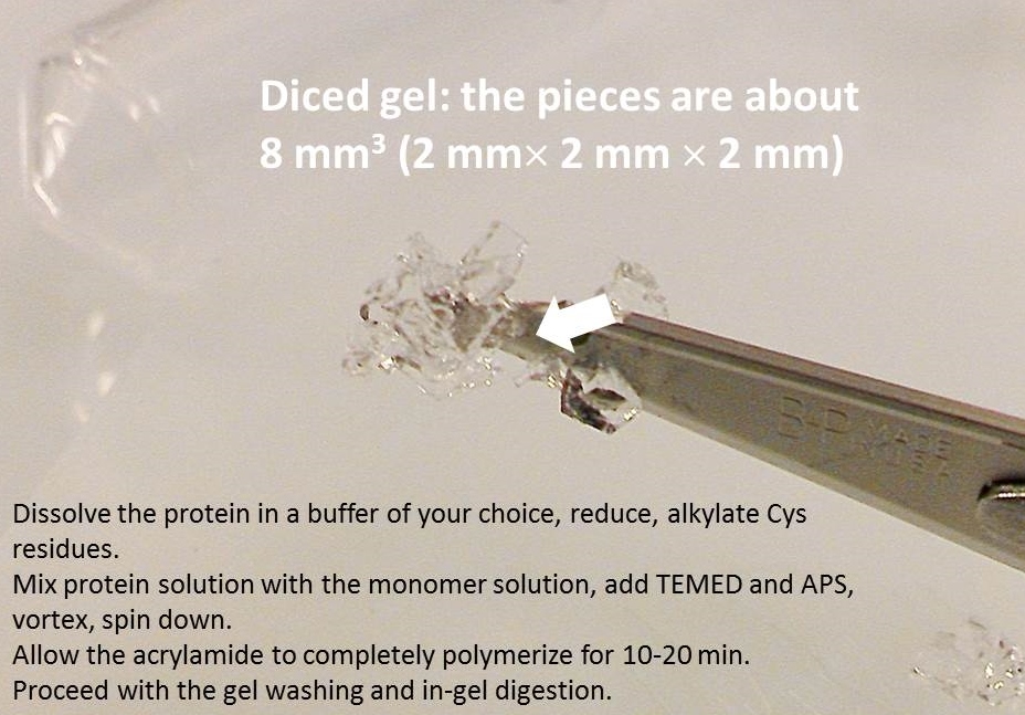
2. Dice the band into 1-mm (1/16”) sections; this will help the protease reach more protein inside the gel.

3. Place the gel cubes in a clean microcentrifuge tube, cover with water to prevent them from drying out, and label the tube with no more than 5 characters. For example, use your initials and numbers.

4. Include a positive control! There is no charge for analyzing a control! Cut out a known protein band following steps 1, 2, and 3.

Cutting a gel band

The in-gel digestion protocol can be found on Thermo website along with the product numbers for all necessary reagents.

Washing the gel

You will need 50% acetonitrile (ACN) solution containing 8 mg/mL ammonium bicarbonate (wash solution). Add 0.05 – 0.1 mL of this solution per gel band, enough to completely cover the gel. Incubate at 37 C for 15-20 min, discard the solution. Repeat two more times. At this point, all or most of the blue color should be gone. If you are destaining SYPRO, there is no easy way to tell whether it is all gone, of course.

Reducing disulfides and alkylating Cys residues

You will need

  1. 5 mM TCEP (Tris(2-carboxyethyl)phosphine hydrochloride) solution in the 8 mg/mL ammonium bicarbonate. I make 100 mM (29 mg/mL) stock solution in water and store it in the fridge up to a month. Dilute this stock 20x (e.g. 50 uL 100 mM TCEP + 950 uL ammonium bicarbonate solution).
  2. Fresh 100 mM IAA (iodoacetamide) solution in the 8 mg/mL ammonium bicarbonate. This is approximately 18 mg/mL IAA solution.

Cover the gel slices with 5 mM TCEP and incubate 10 min at 60 C. Remove the TCEP solution and cover the gel with 100 mM IAA solution. Incubate at 37 C for 15 min with occasional shaking, preferably protected from light. Room temperature will also work, but give it extra 10-15 min. Discard the IAA solution and wash the gel with the wash solution three times to remove IAA and TCEP. (In the Pierce protocol, you might notice that 100 mM IAA requires 9.3 mg in 1 mL, while MW of IAA is 184.9. It is a typo. Either way: 50 mM, 9.3 mg/mL or 100 mM, 18 mg/mL, will work as there will be a large molar excess of IAA compared to Cys. Shrink the gel by covering it with ACN and incubating at r.t. for 15 min or until the gel turns white and brittle. Remove ACN and allow the gel to air dry for 15 min at 37 C (or longer at room temperature).

Digestion

Make 1 mg/mL trypsin stock solution in 50 mM acetic acid or 1 mM hydrochloric acid. This solution can be aliquoted and stored in a freezer for months. Dilute trypsin to 0.01 mg/ml (1:100 dilution) with 8 mg/mL ammonium bicarbonate. Add 50 uL of this solution per gel band and incubate at 37 C 8-24 hours. If you are working with a large piece of gel or several bands combined in one tube, make sure that after the gel re-hydrates in the enzyme solution, it is completely covered. Add more enzyme solution if necessary.

Extracting the peptides

You will need 50% ACN solution containing 0.1% formic acid (FA). If you don’t have formic acid in your lab, let me know – we will share ours with you. Transfer the digest solution to a new tube. Extract each gel band with 50 uL of 50% ACN/0.1% FA (more, if working with a large volume of gel) by incubating at 37 C for 15 min. Transfer this solution to the new tube with the digest solution. Repeat 2 more times. Evaporate the combined extracts in a vacuum concentrator (e.g. Speedvac). Please note, if you are extracting large peptides (>5000 Da), using a sonic bath may be a better option than incubation. Also, in addition to the three 50% ACN extractions, you can use 100% acetonitrile for the final extraction. Submit your dried samples along with a picture of the gel so that I can estimate how much of each sample to use for analysis, and don’t forget to fill out the Protein ID Request form

As always, let me know if you have any questions!

Fingers crossed…

Photo by Evan-Amos

Photo by Evan-Amos

If someone told me “I honestly believe, the instrument is calibrated, it seemed okay last year when I checked it last time. It looks like the mobile phase is a bit old, I am pretty sure I replaced it 3 months ago when a spider fell into the bottle, so it should work. It is recommended to clean the ion source daily, but who has time, let’s hope it is not too dirty. I’m just going to run your samples and see what happens. Fingers crossed!” chances are, I would take my samples to be analyzed elsewhere.

So when I hear “We used old reagents, but they should still work, right? I found this old trypsin in our lab, I hope it is still okay. I am pretty sure the protein is still there, but it might have degraded. We bought new co-IP beads and are unsure how much protein we have loaded, but fingers crossed – everything worked!” I lose my mojo. If crossing fingers is a part of the experimental design, how are we going to troubleshoot when something goes wrong? Was it old trypsin or was the hypothesis no good?

Good news! There is a way to check if the old reagents still work and if the beads have the expected capacity – run a pilot experiment using something that always works; run a control experiment (or two) in parallel with the real experiment. Carry your control through the same procedure as your unknown. Check every step in the protocol: did it work? Our facility loves controls so much that there is no additional charge for analyzing them. So go ahead, bring as many controls as you think are necessary and you won’t have to cross your fingers!


1D and 2D PAGE equipment and supplies

I get a lot of inquiries about our PAGE (polyacrylamide gel electrophoresis) equipment, so here are some information on the equipment we have and the supplies we use.

Cost: It’s free!

We share our PAGE equipment with you at no charge. You need to purchase your own supplies: e.g. gels, standards, buffers, and other consumables. Sometimes we have supplies left over from the Facility experiments, which we will gladly share with you as well. Please, ask before you buy your own!

1D PAGE

Power supply

Our power supply is a basic model; it does not store programmed methods. It can run up to 4 cells simultaneously, as long as the conditions are the same.

Electrophoresis cells

We have three Bio-Rad electrophoresis cells, a Mini-PROTEAN Tetra for up to 4 mini-gels (8.6 x 6.7 cm), a Criterion cell for up to 2 midi-gels (13.3 x 8.7 cm), and a Criterion Dodeca cell for up to 12 midi-gels. We also have a blotter for 13.3 x 8.7 cm gels.

2D PAGE, first dimension (isoelectric focusing, IEF)

Our PROTEAN IEF cell (Bio-Rad) is capable of storing methods, and accepts 7 cm, 11 cm, and 18 cm focusing trays. We also have rehydration trays for all three IPG (immobilized pH gradient) strip lengths.

Gel casting equipment

You can use our gel casting stands and glass plates (0.75 mm) to cast your own mini-gels. In my experience, 1-mm thick gels hold up much better, especially for low %T (4-10%). Bring your own plates if you wish!

Supplies

The Pierce precast mini gels from Fisher are compatible with the Bio-Rad Mini-PROTEAN cell. We do not buy precast gels from Bio-Rad due to the high shipping costs. Fisher orders ship free. You can learn more here.

Immobilized pH gradient (IPG) strips are also available from Fisher (GE Healthcare Immobiline DryStrip gels, for example). The prices are comparable with the Bio-Rad’s, and the shipping is free.

05/28/2014 ETA: According to the GE Healthcare website, Immobiline IPG strips and buffers are not compatible with the Bio-Rad IEF cell running conditions. I am not familiar with the GE system and cannot tell you whether this is true; so to be safe, please use Bio-Rad IPG strips and buffers. If anyone has successfully run Immobiline strips on Bio-Rad IEF cell, please share with us! This information could save your colleagues both time and money!

Disclaimer: We do not receive any form of compensation for promoting any brand or a particular distributor.

Still have questions? Let’s meet and talk about your project!


What is TMT?

TMT (tandem mass tags) are labeling reagents for comparative mass-spectrometry-based proteomics, a Thermo equivalent of iTRAQ. Most popular TMT are amine reactive, but SH-reactive tags are also available. Each isobaric TMT reagent has an amine-reactive NHS-ester group, a spacer arm, and a tandem MS reporter. Either intact proteins or their tryptic digests can be labeled, and up to 6 experimental conditions can be compared in terms of protein expression differences. You can find more information about TMT reagents here.

Our facility is capable of performing both TMT and iTRAQ experiments.

Am I going to get my sample back?

This is a legitimate question! After all, you worked hard to prepare that sample.

The answer is no, your sample is destroyed during the analysis. What happens? Molecules in your sample become ionized, enter the mass spectrometer, and eventually collide with the mass analyzer electrodes. Once a year or so, we open the instrument and clean off the electrodes.

On the bright side, we only need to destroy a small amount of your sample, less than a microgram.