Multi-Method Approach to Biomechanics Research
By: Dr. Ben Infantolino, Assistant Professor of Kinesiology
Biomechanics is the study of biological systems using the principles of mechanics. Since muscle is responsible for producing human motion, it is a very relevant topic of study in Biomechanics. Computer models of muscle are critical to our understanding of muscle function and have been purported to be used to direct surgeries that are designed to improve movement for individuals with a movement disorder. My research focuses on how whole muscle produces force and how variability in muscle can affect the computer model results.
 In my research, I use a variety of methods to examine muscle force production capabilities. I use cadaveric specimens, magnetic resonance imaging (MRI), and ultrasound, as well as live subject protocols to investigate muscle. By using a variety of methods, I can overcome the shortcomings of one method by another.
Cadavers specimens offer unrestricted access to muscle tissue. Anything (besides force) that is of interest in terms of muscle function can be measured on cadaveric muscle tissue. Much of my research has used cadavers to measure basic parameters of muscle (mass, length, angulation). These basic parameters are critical inputs to muscle models.
In many cases, current muscle models combine parameters from different previous studies for the inputs of the muscle model. My research has focused on highlighting the variability that exists between cadavers (and by extension live subjects) for the critical parameters necessary for the construction of a muscle model. I have shown that variability has a large effect on muscle model output. Two areas that cadaver dissection is limited in is the ability to produce force like a live subject, and the inability to easily visualize the three-dimensional architecture of the components that make up a muscle.
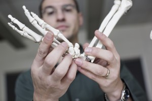
Whole muscle is comprised of fascicles which are bundles of muscle cells. How these fascicles are arranged in a muscle determines the range of motion and force production capabilities of muscle. Since these fascicles exist in three dimensions, traditional dissection cannot be used to determine the arrangement of the fascicles throughout the whole muscle volume.
High field Magnetic Resonance Imaging (14 tesla) MRI can be used to take cross-sectional images, which are then used to reconstruct fascicle arrangement in three dimensions. Fascicles that are arranged in series will produce a muscle that has a large range of motion and can shorten quickly while fascicles that are arranged in parallel produce a muscle with high force production capabilities.
 My research has shown even small muscles have serially arranged fascicles that wrap around a central tendon. The exact function of the fascicle wrapping is unknown. Using high field MRI to view muscle poses two problems: the bore size of the MRI is too small for a live subject to place a limb into the machine, and the scan time is too long (~14 hours) for a subject to remain still in the machine.
Ultrasound is a real-time imaging system that allows for the production of a cross-sectional image of muscle on a live subject while they are moving. These real-time images or videos can be used to measure many of the basic parameters of muscle including muscle volume and fascicle angulation. Ultrasound can be combined with machines that can measure the force exerted by a limb and the limb’s position in space, which allows for the determination of even more parameters of muscle function.
In my research, I have fully characterized the First Dorsal Interosseous muscle noninvasively in live subjects. Unfortunately, to fully characterize a single muscle you need a mathematically determinate system, which is rare in the human body with many muscles crossing a single joint. The solution to this problem is to use cadaveric data to aid in splitting up the contribution of each individual muscle.
I have initiated a healthy undergraduate research program at Penn State Berks. Students who are in Science Emphasis of the Kinesiology program at Berks are required to complete a thesis their senior year. This past year, five students produced publication-level work relating to my research program with one manuscript currently in review.
 Students are now starting to work with me on my research projects earlier than their senior year to gain additional experience before their senior research project. Involvement in undergraduate research provides a strong research foundation for many of our students who continue on to Doctorate of Physical Therapy or other graduate programs.
Muscle is an exciting area of research because it is a challenging tissue to study. Using many different research methods helps elucidate more about muscle than one single approach can. This multi–method approach also allows for many students to be involved in my research, and produces a research plan where one area supports another.