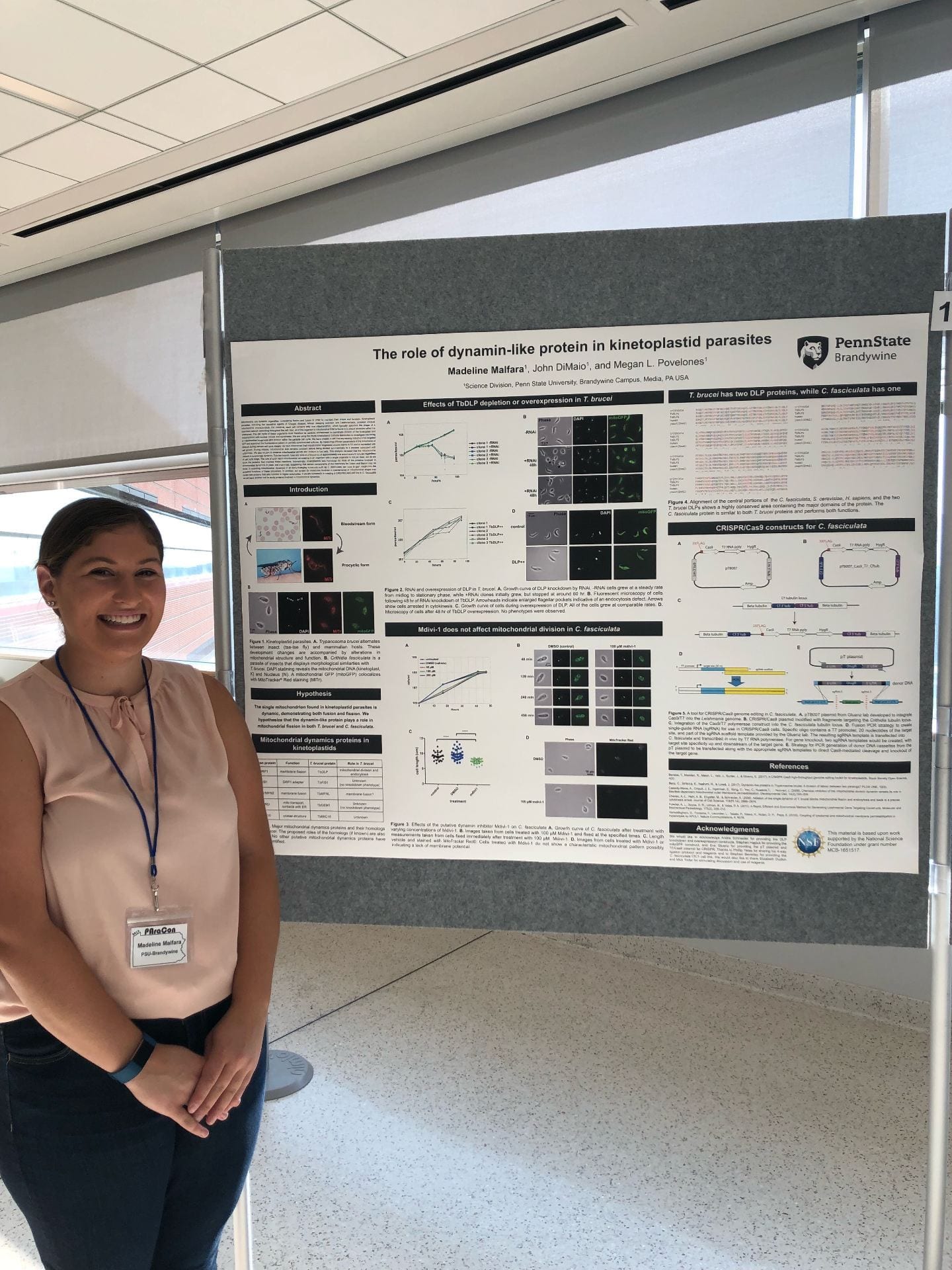
Cells are three-dimensional, a fact that is sometimes hard to appreciate from textbook illustrations. Mitochondria in particular can have complex three-dimensional structures, especially the reticulated mitochondrial networks of kinetoplastid parasites! To illustrate this, we are working with engineers at Penn State Great Valley to print 3-D models of kinetoplastid mitochondria. This is a great collaboration between our lab, Gordon Ruthel at the Penn Vet Imaging Core, Dennis Wozniak, Engineering Lab Manager at Great Valley, Abbey Philip, ITS network/system administrator at Great Valley, and Kathryn Jablokow, Professor of Engineering at Great Valley.
We plan to use these models to address pedagogical questions about how students approach problems in 3D quantitative cell biology, and to communicate our work to colleagues and the general public.
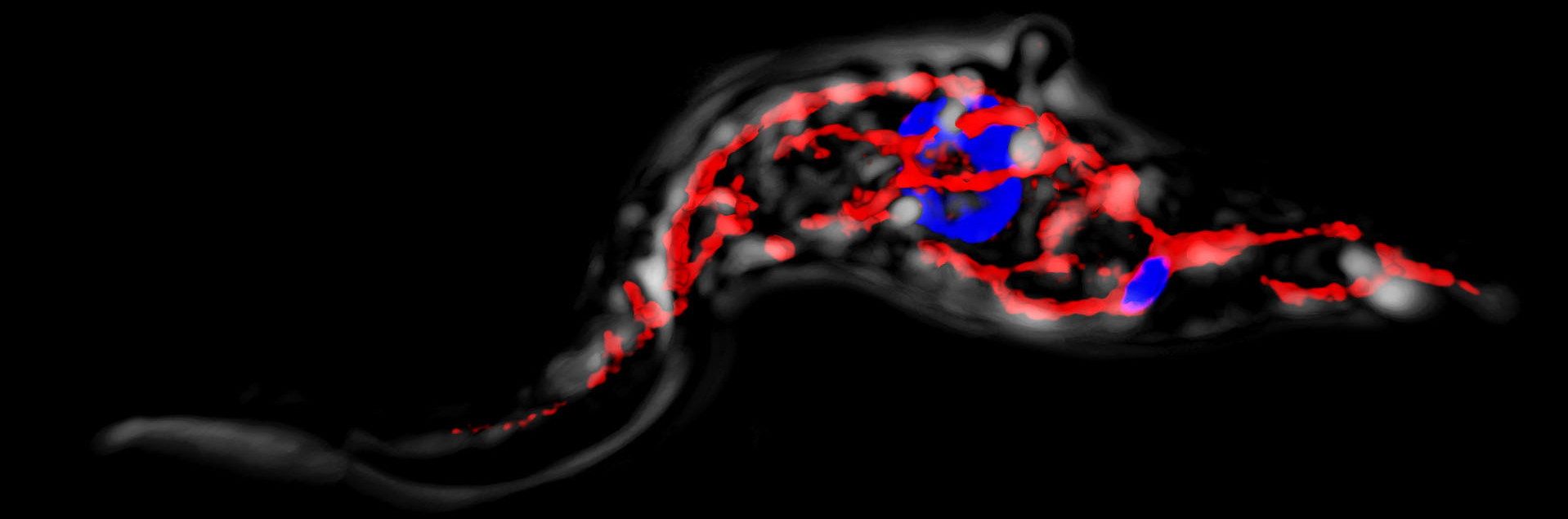
This is one of our first models. It is a G1 (non-dividing) stage Crithidia fasciculata mitochondrion. The original cell expressed mitochondrialy-targeted GFP (mitoGFP), and was fixed and imaged on a confocal microscope. With confocal, we can image the entire cell as a series of thin slices and then assemble the slices into a stack, called a z-stack, in order to obtain 3D information. We were then able to convert that 3D information into a format that was readable by the 3D printer.

Looking at the model I was struck by how differently I interacted with it compared to a 3D rendering I can rotate on a computer screen. I also noticed detailed aspects of the structure I hadn’t appreciated before!