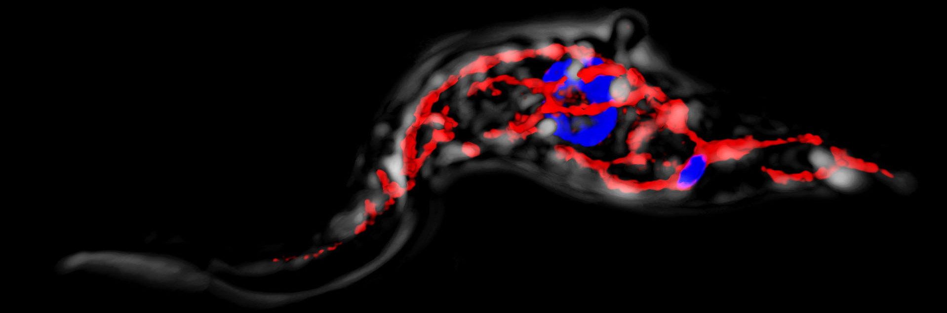
A movie obtained at the Penn Vet Imaging Core (PVIC) of a Crithidia fasciculata cell expressing a mitochondrially-targeted GFP (green). DNA (DAPI) is shown in blue. A skeletonization of the image shows how we can use these 3-D images to quantitate various features of the mitochondrial network.
Crithidia fasciculata colonization of a laboratory strain Anopholes stephensi mosquito.
A time-lapse movie of a C. fasciculata cell expressing mitochondrial GFP imaged on a spinning disk confocal microscope at the PennVet Imaging Core.
