
Radiofrequency Ablation (RFA) is a common minimally invasive cancer treatment modality for patients who are unwilling or unable to undergo a surgical resection of abdominal tumors. RFA is performed by heating the tissue with an electrode inserted into the tumor. The shape of the treatment zone is primarily determined by the shape the electrode and properties of the surrounding tissue. Endoscopic ultrasound-guided radiofrequency ablation (EUS-RFA) offers access to additional regions of the abdomen that were previously inaccessible through traditional percutaneous RFA. Current EUS-RFA electrodes are straight, generating an elliptical ablation zone, and offer limited ability to treat the entire tumor; see Figure 1.
Straight electrodes often require multiple insertions and treatments in order to destroy a single tumor because of the mismatch between the ablation zone and tumor shape. In order to understand RFA, numerous finite element models have developed. While computational models have been developed to study a variety of different aspects of RFA, there have been no attempts to use these models to determine the optimal shape of an RFA electrode for a specific tumor shape.
The goal of this project is to develop a systematic optimization approach for generating EUS-RFA electrode designs that are able to more effectively treat tumor geometries. This project includes the development of a novel deployable EUS-RFA electrode (see Figure 2), optimization of the electrode geometry by coupling an RFA finite element simulation with a genetic algorithm (see Figure 3), and validation of the electrode design and finite element models through ex-vivo and in-vivo experimentation (see Figure 4) [1–3].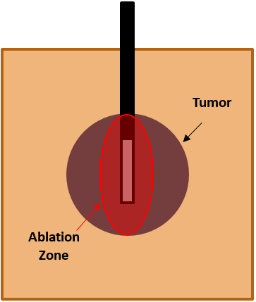
Figure 1: Schematic of Radiofrequency Ablation (RFA) showing the region of treatment, or ablation zone, surrounding an electrode that has been inserted into a tumor. The mismatch between the tumor shape and ablation zone results in the need for multiple electrode insertions or treatments in order to completely destroy to the tumor.

Figure 2: A novel deployable electrode design has been developed that stows inside an endoscopic needle and then deploys into tissue. The design is a circular array of flexible needles, or tines, surrounding a stiff central tine. The electrode shape is manipulated by a set of design variables such as the length (L) and radius of curvature (R) of the outer tines and length (C) of the central tine.
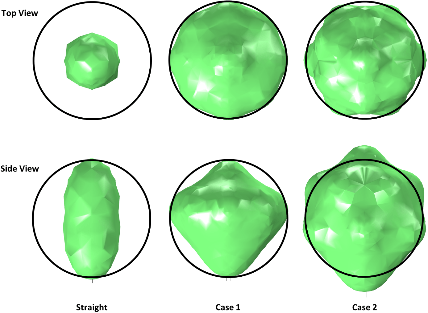
Figure 3 Using a systematic design optimization method that couples an RFA finite element simulation with a genetic algorithm, the electrode geometry has optimized for two electrode variations (Case 1 and Case 2) and compared against the current technology, a straight electrode. The black outlines represent the target tumor shape, a 2.5cm spherical tumor. The green surface represents the ablation zone generated surrounding the electrode. The straight electrode destroys only 25% of the target while Case 1 and Case 2 destroy 71% and 87%, respectively.

Figure 4 The ex-vivo experimentation will be used to validate the RFA finite element model. The electrode is inserted into a thermochromic tissue phantom in which the ablation zone is marked by the color change of the phantom. The tissue phantom is surrounded by a heated saline solution that provides the electrical connection to the grounding pad. After ablation, the tissue phantom is removed and sliced in various cross sections to measure and compare against finite element simulations.
Team Members
Brad Hanks
Fariha Azhar
Katherine Reichert
Dr. Matthew Moyer (M.D.)
Benjamin Kusiak
Project Sponsor
National Science Foundation (NSF)
References
[1] Hanks, B., Frecker, M., and Moyer, M., 2016, “Design of a compliant endoscopic ultrasound-guided radiofrequency ablation probe,” ASME International Design Engineering Technical Conferences and Computers and Information in Engineering Conference, pp. 1–9.
[2] Hanks, B., Frecker, M., and Moyer, M., 2017, “Optimization of a Compliant Endoscopic Radiofrequency Ablation Electrode,” ASME International Design Engineering Technical Conferences and Computers and Information in Engineering Conference, ASME, Cleveland, pp. 1–12.
[3] Hanks, B., Frecker, M., and Moyer, M., 2018, “Optimization of an Endoscopic Radiofrequency Ablation Electrode,” ASME J. Med. Devices, 12(3), pp. 1–11.