How Athletes See: The Primary Visual Pathway

The primary visual pathways in the brain allow athletes to perceive and process visual information. The visual space that healthy athletes can see results from different areas of the brain, or cortical areas, working together to integrate information.

The axons of neurons in the thalamus that travel through the primary visual pathway mediate vision and visual perception. The cortical neurons in the temporal lobe respond to edges in certain orientations or the movement of edges in a particular direction. The primary visual pathway also contains the dorsal and ventral pathways used to process information about faces, shapes, and colors or an object’s relative location and movement.

The Neural Pathway Allowing for Vision

The primary visual pathway is the main neural pathway that allows athletes to see their visual scenes. This pathway’s neural path precedes in the following order: the eye, the thalamus, and the primary visual cortex.

A drawing demonstrating the visual pathway. A horizontal top view of the brain shows the two eyes at the front. Visual field of the left eye sends visual sensations to the right visual cortex through optic nerves and the optic tract. Similarly the right visual field sends information to the left visual cortex. Binocular field information of the left and right eye is sent to the left and right visual cortices respectively. The optic tract connects to the lateral geniculate nucleus of the thalamus. The pituitary gland is labeled and appears at a point centrally behind the two eyes. The optic nerves for each eye intersect behind the pituitary gland, and further behind this is the suprachiasmatic nucleus of the hypothalamus to which branches of the optic tract also connect.

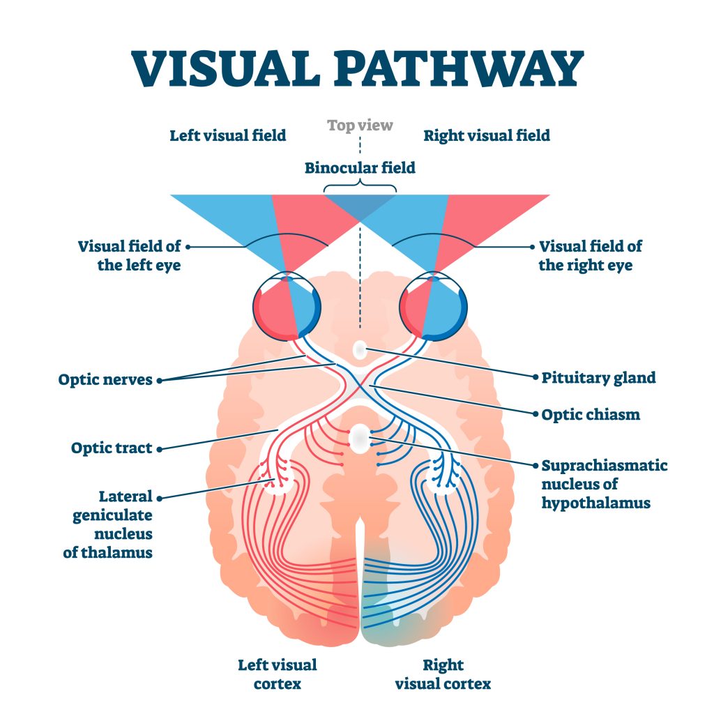
Photo 1: The article Vision, published by Dalhousie University’s Department of Psychology and Neuroscience, displays the primary visual pathway.

Spatiotemporal Tuning of the Neurons in the Visual Cortex Bars and Edges

Cortical neurons in the temporal lobe respond to the orientation of edges seen in the visual space. Each neuron will have a preferred orientation or movement for edges in a particular direction. When the visual space contains this orientation, the neurons send signals to the brain. Therefore, a specific visual scene is encoded by distinct neurons that respond to certain edges of that visual scene.

Photo 2: The textbook Neuroscience by Dale Purves displays how different orientations of the image of the dog produces the image of the dog seen in the visual scene.

Dorsal and Ventral Divisions in the Primary Visual Pathway

The thalamus is separated into two layers: the magnocellular layer and the parvocellular layer.

Layer of Thalamus Cell Type Convey Information About
Magnocellular Layer M Cell  Motion
Parvocellular Layer P cells Color

A study titled “Interactions Between Dorsal and Ventral Streams for Controlling Skilled Grasp” by Vonnevan Polanen found that the connections between these two layers of the thalamus are important for controlling complex object-oriented hand movements in addition to allowing for perception of the visual scene.

Organization of the Extrastriate Visual Areas

The extrastriate visual areas of the brain process other information about a spatial scene. It is organized into the dorsal pathway and ventral pathway.

  • Ventral Pathway: The perception of faces, colors, and shapes
  • Dorsal Pathway: The location of objects and their movements

Photo 3: The textbook Neuroscience illustrates that the ventral pathway involves the brain’s lower areas. The dorsal pathway involves the brain’s upper areas.

Injuring a Critical Core Muscle: Quadratus Lumborum Syndrome

The quadratus lumborum is a back muscle that athletes utilize daily for balance and stabilization. The downside of the muscle playing a crucial role in supporting the body is that it can easily be injured. Aching pain in the lower back and tenderness characterizes the diagnosis of quadratus lumborum syndrome. The mechanisms of injury are linked to trauma or repetitive daily activities. The treatment options for the syndrome include ultrasounds, medications, or physical therapy.

What is the Quadratus Lumborum

An article by Krzysztof Kassolik titled “Quadratus Lumborum Syndrome” states the quadratus lumborum is the deepest muscle in the back. The muscle starts on the bony protrusion of a vertebra in the lower back and ends on the pelvic bone.

Photo 1: An article titled “Myofascial Lower Back Pain” by Ryan Ramsook illustrates the quadratus lumborum’s placement on the ribs and pelvic bone.

Diagnosing Quadratus Lumborum Syndrome

Quadratus lumborum syndrome characteristics include the following:

  • Muscular tenderness
  • Muscular twitching responses

Quadratus lumborum syndrome can cause pain during the following motions or positions:

  • Deep aching pain persisting in the lower back in a sitting or standing position
  • Sneezing or coughing
  • Lying down or walking up stairs

Mechanism of Injury

The mechanism of injury for quadratus lumborum syndrome can be from repeated use or a traumatic injury.

  1. Traumatic injuries: Traumatic injuries affecting the quadratus lumborum muscle include car accidents or sports injuries.
  2. Repetitive Daily Activities: Lifting a heavy object can strain the quadratus lumborum.
  3. Sitting for Long Periods: When an individual is sitting for too long, this often causes continuous contraction and tightening of the quadratus lumborum muscle. When the muscle is continuously contracting, it can lead to fatigue. This sitting position can also lead to decreased blood flow to the muscle, leading to stiffness and pain.
  4. Weak Back Muscles: Weak muscles around the quadratus lumborum can also cause pain in the muscle. When the surrounding muscles in the back and pelvis areas are too weak, the quadratus lumborum may have to work hard to support the body. Eventually, after working too hard when sitting and standing, the quadratus lumborum may become overworked and tense.

Photo 2: An article by Rick Perry of BQ Personal Training titled “Poor Posture: The Root of Neck & Back Pain” illustrates areas of back pain based on the type of posture an individual has.

Treatment

Quadratus lumborum syndrome can be treated with non-surgical methods or with physical therapy.

  1. Ultrasound Infiltrations: Ultrasound-guided infiltrations of the quadratus lumborum are an effective procedure to relieve pain in the quadratus lumborum syndrome.
  2. Medication: Taking non-steroidal anti-inflammatory drugs, opioids, anticonvulsants, antidepressants, and muscle relaxants is a non-surgical treatment option. Anticonvulsants can block the pain coming from the quadratus lumborum by blocking the sodium and calcium channels at a cellular level, suppressing the continuous muscle contractions in the muscles.
  3. Physical Therapy: Physical therapy can also alleviate the point-tenderness of the quadratus lumborum with the use of exercise treatment plans and manual therapy.

Nitric Oxide’s Importance in Analyzing Microvascular Function

Nitric oxide is released by endothelial cells of blood vessels when the skin is heated. Protocols have been performed where the skin is heated to 39°C to determine nitric oxide contribution to vasodilation and the resulting increase in blood flow. Clinically, this protocol can test for microvascular function in older individuals or those in disease states.

Exercise Caused Heating of the Skin

When athletes are exercising, their skin can often appear red. This redness is due to vasodilation occurring in blood vessels. Vasodilation occurs when endothelial cells of blood vessels relax their smooth muscle lining and increase in diameter. This vasodilation increases blood flow to reduce the body’s core temperature.

Nitric Oxide’s Contribution to Heating the Skin

The smooth muscle relaxation is partly due to the vasodilator nitric oxide (NO). NO is produced in endothelial cells by the amino acid L-arginine using the nitric synthase (NOS) enzyme.

A study done by Patricia J. Choi at the Department of Human Physiology, University of Oregon, titled “A New Approach to Measure Cutaneous Microvascular Function” found that heating the skin to 39°C increases skin blood flow and vasodilation. In this study, the pharmacological agent l-arginine methyl ester (L-NAME) was perfused into the skin to measure nitric oxide contributions during vasodilation.  This perfusion method via microdialysis was paired with laser Doppler flowmetry to measure skin blood flow.

figure 5

Photo 1: The European Journal of Applied Physiology article by David Low titled “Assessment of Human Cardiovascular Function” depicts the microdialysis and the laser Doppler flowmetry technique.

The Importance of Utilizing Nitric Oxide for Skin Heating

Measuring the release of nitric oxide using a local heating test and L-NAME can be useful for measuring the microvascular function of endothelial cells. NO availability underlies many diseases and conditions of older individuals. Therefore, testing for the NO-dependent microvascular function can be clinically valuable.