Energy-dispersive X-ray spectroscopy (EDS) can be used to examine the elemental composition of of a sample. Flor used EDS to map the presence of zinc in the mandibles of fall armyworms, Spodoptera frugiperda, showing that the metal is accumulated at the edges of the mandibles to strengthen them (take a look at her blog post here).
As part of my work on the semitransparent patches found in Ceraphronoidea, I wanted to use EDS to see if there were any differences between the semitransparent patches and the surrounding cuticle (for an overview of what the semitransparent patch is, take a look at my blog post from István’s “Know your Insect” seminar).

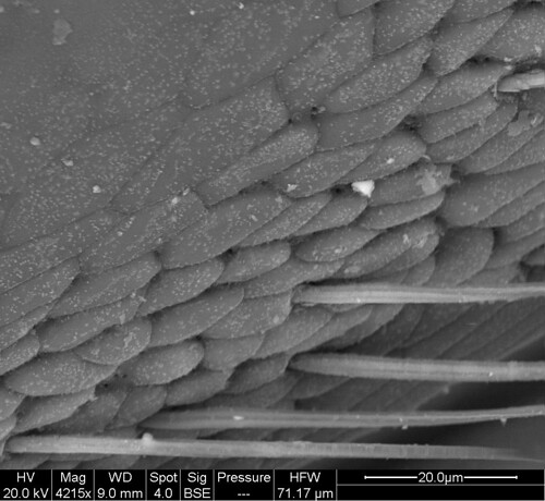
While using EDS to explore the semitransparent patches in Megaspilus armatus, we were able to take some nice SEM (scanning electron microscope) images. I was surprised at how scaly the nearby felt field looked. When we zoomed in, we were able to clearly see the pores that I found during TEM and SBFSEM.
Though our initial EDS work didn’t seem to show a difference in the elemental composition between the semitransparent patches and the surrounding cuticle, there might be a concentration of calcium, sodium and phosphorus around the felt fields. Could these elements be components of the substances secreted from the pores? I look forward to analyzing my data so I can find out!
Leave a Reply