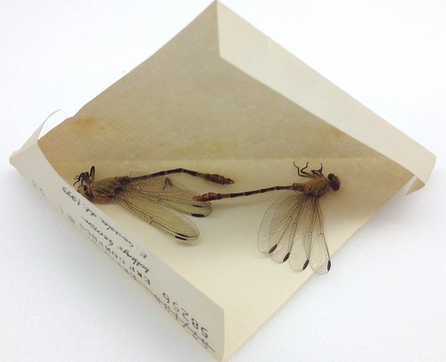
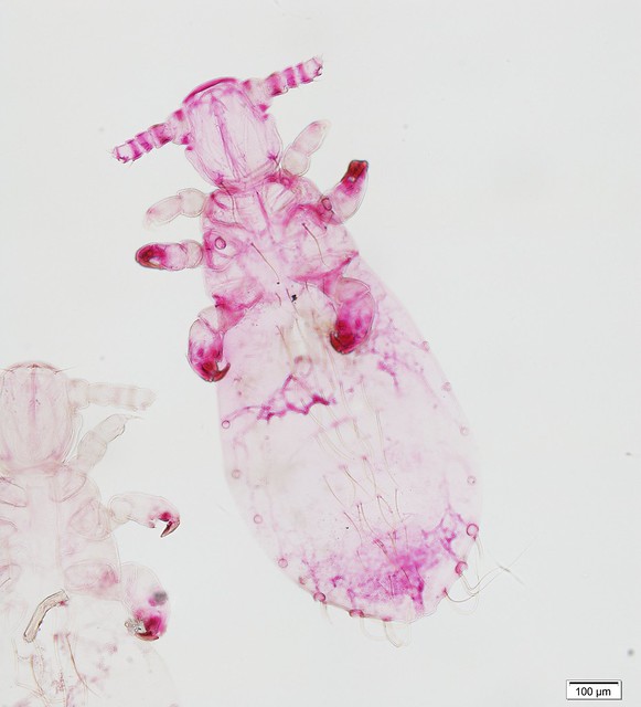
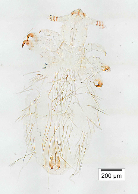
This week’s Friday Night Lice mystery louse parasitizes a host from an order of mammals we haven’t yet covered in this series. So, it’s not found on rodents, carnivores, pinnipeds, primates … Can you guess what it is? It was collected in Nepal in the late 1960s. Offer your guesses as comments below! We’ll post the answer on Monday.
Research Symbiosis: Anjel Helms and the rectal glands of tephritids
Anjel Helms is a PhD candidate in the Tooker Lab here at Penn State, with an impressive array of academic accomplishments (see Google Scholar). It is a real pleasure for us at the Frost to collaborate with Anjel on a project concerning rectal glands (see smartbox) of the Goldenrod Gall Fly (Diptera: Tephritidae: Eurosta solidaginis).
Smartbox 1. Rectal glandsAnimals that eliminate their nitrogenous waste in the form of uric acid (e.g., birds and reptiles), show an active reabsorption of water from the excrement in the cloaca. The continuous circulation through the excretory system enables these animals to accomplish their excretion with a minimal amount of fluid. In mammals, the water secreted in the digestive juices is reabsorbed, resulting in more or less dry feces before they leave the body. Similar circulation of water takes place in insects, where much of the reabsorption occurs in the lower segments of the Malpighian tubes, but some takes place also in the rectum. The sites of reabsorption in the rectum are the so called “rectal pads”, where the wall of the rectum is thin and lacks musculature (Wigglesworth 1932). In some insects, these pads invaginate inside the lumen of the rectum, forming the rectal papillae. In insects with diluted rectal content, including some dipterans, lepidopterans and siphonapterans, the rectum does not play a role in water reabsorption and, in some cases, has evolved a different function (Wigglesworth 1965). For example, in males of some tephritid species, the rectum is extended into a large reservoir, called the “rectal gland”. This reservoir stores pheromone compounds that increase the sexual receptiveness of females. The rectal gland reservoir enlarges gradually during the maturation of the male specimens, and is capable of storing a substantial volume of liquid pheromone (Lee and Chang 1986). The stored compounds are then eventually released, presumably due to the contraction of the two muscle layers in the wall of the reservoir (Kho and Tan 2005). The origin of the pheromone compounds stored in the rectal gland is largely unknown. A small, cone-like evagination at the distal end of the rectal gland, the “secretory sac” is suspected to produce pheromone compounds (Fletcher 1969, Schultz and Boush 1971). This hypothesis is supported by the fact that the secretory sac is absent from females. Studies focusing on one tephritid species, the asian papaya fruit fly (Bactrocera papayae) show that the most effective compounds of the pheromone (phenylpropanoids that were biotransformed from host-plant-derived methyl eugenol) are transported from the hemolymph to the rectal gland via the rectal pads (Kho and Tan 2005). The main components of the pheromone blend of the goldenrod gall fly (E. solidaginis), as well as a few other tephritid species, are spiroacetals. The biosynthesis of insect spiroacetals is not well understood, but it has been proposed that these compounds may be derived from fatty acids (Francke and Kitching 2001, Booth et al. 2009). |
My interest in the goldenrod gall fly (Eurosta solidaginis) began when I arrived at Penn State in August 2010. My adviser Dr. John Tooker had been working with these flies for a few years already and his enthusiasm for them (and gall insects in general) was contagious. While conducting research on the goldenrod gall fly, John found that the male flies emit very large quantities of a volatile emission (~70 ± 20 μg over 24 h) that is attractive to female flies (Helms et al. 2013). In spring, adult males perch on goldenrod plants and emit pheromone to attract potential mates. We were intrigued by the flies’ substantial pheromone production (many lepidopteran species emit only a few nanograms of pheromone over the same time scale) not only because of the resource allocation required to produce the compounds in the emission, but also because of the strong environmental signal it provided. Dr. Mark Mescher, my co-adviser, is interested in the ecology and evolution of communication systems, especially chemical communication involving plants and insects. The combined research interests of my two advisers led me to develop research questions centered around chemical communication and defense between E. solidaginis and its host plant, tall goldenrod (Solidago altissima). My primary research goal was to test whether goldenrod plants eavesdrop on goldenrod gall flies by perceiving and responding to the male pheromone emission.
Previous research has shown us that olfactory cues play a central role in many ecological interactions, including those among plants and insects. Some well-documented examples of these interactions include pheromonal communication among insects and the use of plant odors as foraging cues by insect pollinators, herbivores and predators. More recent work has also demonstrated that plants themselves can perceive and respond to olfactory cues. For example, parasitic plants in the genus Cuscuta grow toward host-plant odors and some plant species ready their defenses against herbivores in response to volatile cues emitted by their insect-damaged neighbors. Through our research, we demonstrated for the first time that plants can perceive and respond to olfactory cues emitted by insects. Our findings suggest that tall goldenrod plants exhibit enhanced defense responses following exposure to the pheromone of their specialist herbivore, the goldenrod gall fly (Helms et al. 2013, Helms et al. 2014).
These findings led us to question why male E. solidaginis emit such large quantities of pheromone if it provides a chemical signal for eavesdropping by goldenrod plants. The focus of my current research is examining correlations between male quality (especially related to attractiveness to female flies) and pheromone production. This project led me to contact Drs. Andy Deans and István Mikó at the Frost Museum for help identifying the source of the E. solidaginis pheromone.
In our initial experiments, we collected the volatile emissions of male flies by placing them inside small glass chambers and pulling clean air through the chambers over adsorbent volatile trap filters. We then eluted these filters with solvent and analyzed the fly emissions using gas chromatography coupled with either a mass spectrometer or flame-ionization detector. We were curious where these compounds are produced and stored, and whether the volatile compounds emitted by the flies match those stored inside them. Other studies with tephritids indicated that our flies may utilize a rectal gland for pheromone production and/or storage. István agreed to help me look for the pheromone source, so we started dissecting flies and trying to locate a rectal gland.
At the Frost, we dissected the golderod gall fly specimens in 0.1 M phosphate buffer on lower magnification (80× with our Olympus SZX16 stereomicroscope). We were looking for a structure resembling the rectal gland reservoir, (i.e. an organ the size of a testis, with the distinct rectal pads) such as in Figure 1. in the Smart box. Despite our best efforts, in our first dissection we weren’t able to locate the gland. We were a bit confused and I mentioned to István I thought the gland must be fairly large because when I handled that male, like many others previously, he released a large droplet of liquid that smelled like the pheromone. István then had the idea to carefully avoid disturbing the next male so that he wouldn’t expel any liquid before we dissected him. And sure enough it worked! István was able to locate what looked like a very large rectal gland, complete with rectal pads. It seems the first fly had expelled the contents of his rectal gland, making it small and difficult to locate.
We found a large vesicle-like enlargement on the gut (rec: Figure 3, note that rec on Figure 3 is of a male specimen that has emptied some of the liquid contents while we were macerating; males without emptying the substance have 2-3 times as large rec) distal to the Malphigian tubes). Once we moved to a higher magnification (230×) we were able to identify the rectal pads – with elongate tracheae arising from the middle – on the extended part of the hind gut. This is evidence that the large extension of the hind gut is indeed the rectal gland of the goldenrod gall fly. We also were able to locate a smaller evagination distal of the rectal gland (ext: Fig. 3) that might be equivalent to the “secretory sac” of other tephritids.
We hypothesize that the gland we located is a storage reservoir for the pheromone, which we plan to test by extracting and analyzing its contents. We also predict that the gland contains a secretory sac where pheromone synthesis occurs, but further examination and experiments are needed to confirm this.
We are especially excited to locate the rectal gland of the goldenrod gall fly because this fly’s biology and ecology have been fairly well studied, but its pheromone and rectal gland have received very little attention. The size of the gland was quite surprising for us as it appeared to occupy a significant portion of the abdominal cavity. Based on this discovery, I plan to examine whether a correlation exists between gland size and pheromone production in male E. solidaginis. We also hope the contents of the gland may provide us with a new source of pheromone extract to use in future experiments and that this collaboration with the Frost Museum may lead to future studies of the E. solidaginis rectal gland, including histological and transcriptomic work.
References:
- Booth, YK, Kitching, W, and De Voss, JJ. 2009. Biosynthesis of insect spiroacetals. Nat. Prod. Rep. 26: 490–525.
- Dodson, G. 1978. Morphology of the reproductive system in Anastrepha suspensa (Loew) and notes on related species. Florida Entomologist 61: 231-239.
- Fletcher, BS. 1968. Storage and release of a sex pheromone by the Queensland fruit fly, Dacus tryoni (Diptera: Trypetidae). Nature 219: 631-632.
- Fletcher, BS. 1969. The structure and function of the sex pheromone glands of the male Queensland fruit fly, Dacus tryoni. J. Insect Physiol. 15: 1309-1322.
- Francke, W, and Kitching, W. 2001. Spiroacetals in Insects. Curr. Org. Chem. 5: 233–251.
- Helms, AM, De Moraes, CM, Tooker, JF, and Mesher MC. 2013. Exposure of Solidago altissima plants to volatile emissions of an insect antagonist (Eurosta solidaginis) deters subsequent herbivory. Proc. Natl. Acad. Sci. USA. 110: 199–204.
- Helms, AM, De Moraes, CM, Mescher, MC, and Tooker, JF. 2014. The volatile emission of Eurosta solidaginis primes herbivore-induced volatile production in Solidago altissima and does not directly deter insect feeding. BMC Plant Biology. 14: 173.
- Kho, CC, and Tan, KH. 2005. Rectal gland of Bactrocera papayae: ultrastructure, anatomy, and sequestration of autofluorescent compounds upon methyl eugenol consumption by the male fruit fly. Microsc. Res. Tech. 65: 219-226.
- Lee, LWY, and Chang, TH. 1986. Morphology of sex-pheromone gland in male oriental fruit fly and its suspected mechanism of pheromone release. Fruit flies : proceedings of the Second International Symposium, 16-21
- Schultz, GA, and Boush, MG. 1971. Suspected sex pheromone glands in three economically important species of Dacus. J. Econ. Ent. 64: 347-349.
- Wigglesworth, VB. 1932. On the function of the so-called ‘rectal glands’ of insects. Quart J. Micr. Sci. 75: 131–150.
- Wigglesworth, VB. 1965. The Principles of Insect Physiology. London: Mathuen and Co. Ltd., p 741.
Odes on the verge of royal treatment
Our collection grant is active and spendable, so now’s the time to get serious about our objectives. The biggest projects will happen in the fall and over the holiday break in December/January, but we’ve already started fixing, digitizing, and otherwise preparing the Beatty collection for its new digs. Our draft (emphasis on the word draft, at least at the time of this writing!) workflow is available as a Standard Operating Procedure (SOP) document: draft Odonata SOP
Please comment on this proposed process, especially if you have expertise in Odonata and/or digitizing and/or insect curation in general. Our goals are:
- Image each specimen with its original collecting event label and a unique identifier label.
- Extract specimens from their temporary field envelopes (see image above) and rehouse them in clear cellophane envelopes.
- Move all specimens into acid-free storage, inside sealed, purpose-built cabinets. (No more cigar boxes on open shelves!)
- Convert all photographs to digital records (through crowd-sourcing, probably).
I’ve already had one odonatologist tell me: “Photographing specimens would … be faster than scanning. Still, I think it is a waste of resources. Our funding world might be sufficiently Alice-in-Wonderland that money would be available for this purpose! The taxpayer in me quivers a bit …” That was before seeing our test image (stitch errors and all; see original 16000 x 10000 image). I’m still committed to imaging all specimens, as it provides—minimally—a source for error checking. Thoughts?
Friday Night Lice – week 16 answer
Last week’s Friday Night lice mystery louse was … (drum roll) … Lemurphthirus stigmosus Ferris, 1954 (Polyplacidae). According to its label, the louse in our photograph, specimen PSUC_FEM 10005440-1, was collected in southern Angola in 1954, off of a Senegal Galago, a.k.a. Lesser Bushbaby (Primates: Galagidae: Galago senegalensis). The stated host species looks like this:
According to Wilson & Reeder (2005), however, this species of bushbaby doesn’t live in Angola! My hypothesis, therefore, is that our louse specimen was actually collected from a Mohol Bushbaby (Galago moholi), which is also listed by Durden & Musser (1994) as a host species for Lemurphthirus stigmosus. Here it is:
And another ridiculous photo of a ridiculously cute primate:
Friday Night Lice – week 16
Happy Independence Day to all our U.S. readers! This week’s mystery louse yields few clues as to its biology, as it is a nymph, and it has nothing to do with American independence—at least as far as I can tell. Maybe the fact that the slide is red and white (no blue, though) makes it mildly patriotic?
The specimen was collected in Angola in the 1950s if you need a relatively uninformative hint. No obvious paratergites, smallish grabby bits, sparse coat of setae … Can you guess its host mammal? Another, perhaps more generous hint: unlike most Anoplura this louse species is not a specialist of rodents, but the mammalian host is relatively small. Post your guesses as comments, and we’ll provide the answer on Monday!
- « Previous Page
- 1
- …
- 39
- 40
- 41
- 42
- 43
- …
- 62
- Next Page »