We really enjoyed our recently published research (Mikó et al. 2013) on hymenopteran male genitalia muscles, visualized with the confocal laser scanning microscope (CLSM) at the Cellular and Molecular Imaging Facility at NC State (our previous institution). One of the side projects was this marvelous anatomical system of Megalyra fascipennis (for volume rendered CLSM media files click these Figshare links: 1, 2, 3, 4).
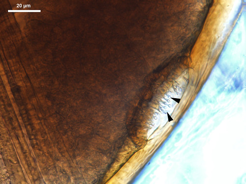
While focusing primarily on M. fascipennis male genitalia, we diverged occasionally onto the rest of the metasoma, eventually discovering two concave impressions on the front edge of the second sternite (S2) with some pore-like openings (Fig. 1).

This structure resembles the cuticular modifications around sternal gland openings (orifices) of termites (Noirot and Quennedey 1974). In termites the cuticle is perforated above some glands, allowing the gland secretion to reach the body surface (in other cases pores are absent so the extract diffuses through the uninterrupted cuticle).
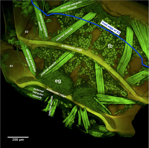
The volume rendered images of the Megalyra specimen revealed that indeed, there is a pair of exocrine glands (‘eg’ in the image below) on the front of the second sternite (S2). Beside the gland we were able to visualize other anatomical structures; the bottom of the metasoma is covered with fat bodies (fb). These organs are analogous with the human liver as they play an essential role in energy storage and utilization.

The fiber-like anatomical structures on the image above are muscles that move the sternites relative to each other (see smart box below).
| Box 1. How Insects are telescoping their abdomen? Sternites and tergites are stiff and harder plates covering the upper- and lower sides of the metasoma. They are connected to each other by more flexible, membrane-like structures (conjuntivae) and muscles. Plates overlap the ones behind them externally, so the front end of each plates is obscured by the plate ahead (in internal view as shown on Figure 2, the plates behind overlap the frontal ones). Plates on the hymenopteran metasoma are moved relative to each resulting the metasoma to act like a small, and very flexible telescope as you can see in this video. This movement is actually caused by the contraction of metasomal muscles. Muscles, in general when contract (shorten) can pull things together but when they relax (elongate) they don’t push them away. For that an extra set of muscles are needed. Three pairs of muscles connect two subsequent plates in the metasoma (image above). Two of these, internal muscle and external protractor muscle connect the front edge of the plates while one pair, the external retractor muscle connects the anterior edge of the plate in the back with the hind part of the plate in the front. Alternate contraction/relaxation of these muscles are mostly responsible for the versatile movement of the metasoma. |
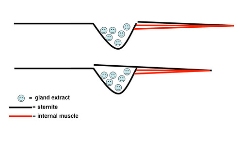
The newly discovered gland of Megalyra is an anterior sternal gland (Buckingham and Sharkey 1988, Quicke 1990) that opens at the front margin of the sternite. Depending of the actual position of the plates, the impression with gland openings are either obscured by the more frontal plate or are disclosed. The telescoping movement of metasomal sternite hence are actually control the release of the gland extract that accumulates possibly in the impression (image below).

This intricate control of gland release is an interesting type of evolutionary adaptation, whereby an existing system serves a new function. Although accurate examination with transmission electron microscope is required for the proper characterization of this new gland we think that it is a nice addition to our knowledge on the Hymenoptera exocrine gland system. Especially that this is the first report on the presence of an exocrine grant from this superfamily.
