Category Archives: 2nd Post 2018

Optimizing the structure of hcp Hafnium

The aim of this project is to determine the unit cell volume using density functional theory (DFT). This work takes advantage of the geometric optimization feature of CASTEP[1], a commercially available plane-wave DFT code. Some details of the results follow, as well as comparisons with previous findings.

Initial conditions and setup

Geometry optimization in CASTEP minimizes the total energy of the unit cell that is provided while allowing variations in the geometry of the provided cell. In the current calculation, we provided the hcp structure for Hafnium pictured below.

Initial cell provided for CASTEP calculation.

The initial values of the lattice parameters are a=b=3.1956 Å and c=5.0511 Å. These values are provided by examples within Materials Studio and are accepted experimental values [2]. We maintain Throughout the calculation, symmetry demands that a=b, but a and c are allowed to vary.

Calculation

The following settings were used for Geometry Optimization in CASTEP:

Atomic calculation performed for Hf:
1s2 2s2 2p6 3s2 3p6 3d10 4s2 4p6 4d10 4f14 5s2 5p6 5d2 6s2

Max. force: .05 eV/Å

Cell optimization: Full

Energy cutoff: 435.4 eV

k-points: 9x9x6 (36 points in the irreducible part of the Brillouin zone (IBZ))

Pseudopotentials: OTFG ultrasoft

Functionals: GGA and PBE

Both the number of k-points and the energy cutoff were varied to ensure reasonable convergence. The values above gave a well-converged energy with a reasonable runtime.

Results

The minimized energy was found to be -15.73336 keV and the corresponding lattice constants were found to be a=b=3.2117 Å and c=5.0557 Å. The unit cell volume was found to be 45.163544 Å^3. This is found to be well in agreement with a result obtained in a previous post [3]. Furthermore, it is known that the ideal packing ratio for spheres arranged in an hcp lattice is c/a≈1.633. In this work, the packing ratio was found to be 1.574, approximately a 4% difference.

References

[1] Clark, S. et al. First principles methods using CASTEP. Z. Kristallogr. 220, 567–570 (2005).

[2] https://www.webelements.com/hafnium/crystal_structure.html

[3] https://sites.psu.edu/dftap/2018/02/14/determining-the-lattice-constants-for-hf/


How initial estimates change convergence properties of bisection method and Newton’s method

Method Introduction

There are various methods that could search local minimums for a function. This locally lowest value search could be called numerical optimization, which could happen in one dimension or  more than one dimension. Normally, the later one is more difficult.

Here, only optimization in one dimension is studied and methods adopted are bisection method and Newton’s method.

These two methods are iterative and can only provide approximations, which is controlled by the convergence criterion. An initial estimate is needed to use these two methods and no guidance is provided by the methods themselves. So the following content will study how the initial guess changes convergence properties of these two methods.

The function used here is f(x)=exp(-x)cos(x). Ranges selected for bisection method are (1.2, 2.4), (1.4, 2.6), (1.6, 2.8), (1.8, 3.0), (2.0, 3.2), (2.2, 3.4). Initial estimates are boundary values of each range for bisection method. Initial estimates selected for Newton’s method are 1.2, 1.4, 1.6, 1.8, 2.0, 2.2, 2.4, 2.6, 2.8, 3.0, 3.2, 3.4. The initial estimates of Newton’s method are consistent with that of bisection method, which is designed for comparison between these two methods.

A reference local minimum for the studied function is x=2.356194490… . How many decimal places is determined by the precision we set. The convergence criterion used here is 0.000001(1E-06). The range selection for bisection is stopped at (2.2, 3.4) (the change step is 0.2 between each range) otherwise the reference value will fall outside the range.

Another tricky thing is that bisection method has two initial estimates, which form a range for later iteration, while Newton’s method only needs one. So, when we doing comparison, choosing which value for Newton’s method should be paid attention. Since Newton’s method converges to different local minimums with bisection method at 3.0, 3.2, 3.4, smaller boundary values of each range(1.2, 1.4, 1.6, 1.8, 2.0, 2.2) are adopted for Newton’s method when we comparing these two methods at each range.

Calculation Results

The difference of each step ε is defined as x value is this iteration minus x value in last iteration.

Following figures show the calculation results. Full data could be got through the link attach at the end of this post.

Comparison for convergence property between bisection and newton’s method

Range (1.2, 2.4)

Range (1.2, 2.4) is chosen for bisection method, the local minimum is 2.356194.

For New’s method, 1.2 is the initial estimate. The local minimum is  2.356194.

Fig 1 shows the convergence properties of bisection method and Newton’s method. Fig 2 shows a part of Fig1 in order to show the convergence detail.

 Fig1. difference of each step ε vs iteration steps at range (1.2, 2.4)


Fig2. part of Fig1 in order to show details of convergence

Both of the methods converge to the same local minimum under the convergence criterion of 1E-06. But the Newton’s method converges needing less steps. The same pattern could be found for following ranges.

Range (1.4, 2.6)

Range (1.4, 2.6) is chosen for bisection method. The local minimum is 2.356194.

For Newton’s method, the initial estimate is 1.4. The local minimum is 2.356194.

Fig 3 and 4 show the convergence properties of the two methods.

Fig 3. difference of each step ε vs iteration steps at range (1.4, 2.6)


Fig 4. part of Fig3 in order to show details of convergence

Range (1.6, 2.8)

Range (1.6, 2.8) is chosen for bisection method. The local minimum is 2.356194.

For Newton’s method, the initial estimate is 1.6. The local minimum is 2.356194.

Fig 5 and 6 show the convergence properties of the two methods.

Fig 5. difference of each step ε vs iteration steps at range (1.6, 2.8)


Fig 6. part of Fig5 in order to show details of convergence

Range (1.8, 3.0)

Range (1.8, 3.0) is chosen for bisection method. The local minimum is 2.356194.

For Newton’s method, the initial estimate is 1.8. The local minimum is 2.356194.

Fig 7 and 8 show the convergence properties of the two methods.

Fig 7. difference of each step ε vs iteration steps at range (1.8, 3.0)

Fig 8. part of Fig 7 in order to show details of convergence

Range (2.0, 3.2)

Range (2.0, 3.2) is chosen for bisection method. The local minimum is 2.356194.

For Newton’s method, the initial estimate is 2.0. The local minimum is 2.356194.

Fig 9 and 10 show the convergence properties of the two methods.

Fig 9. difference of each step ε vs iteration steps at range (2.0, 3.2)

Fig 10. part of Fig 9 in order to show details of convergence

Range (2.2, 3.4)

Range (2.2, 3.4) is chosen for bisection method. The local minimum is 2.356194.

For Newton’s method, the initial estimate is 2.2. The local minimum is 2.356194.

Fig 11 and 12 show the convergence properties of the two methods.

Fig 11. difference of each step ε vs iteration steps at range (2.2, 3.4)


Fig 12. part of Fig 11 in order to show details of convergence


Study for convergence property for bisection and Newton’s method, respectively

The Bisection method

Fig 13 shows the convergence property of bisection method at different range. When only one local minimum exits in the ranges, we can say that optimization in different ranges has the same convergence path, which could be confirmed from data in attachment.

Fig 13. difference of each step ε vs iteration steps for bisection method at different ranges

Newton’s method

Besides 1.2, 1.4, 1.6, 1.8, 2.0, 2.2, Newton’s method could get the same local minimum 2.356194 at 2.4, 2.6, 2.8 for the initial estimate.So the new initial guesses are included for the comparison, which is shown in Fig 14. Only around the local minimum, we can say that the convergence is faster if the initial estimate is closer to the local minimum. When the initial estimate is far from the local minimum, the argument is not right. For example, the convergence is faster when the initial estimate is 1.2 compared with the situation when the initial estimate is 2.8, while 2.8 is closer to 2.356194. The explanation could be that the Newton’s method uses the tangent of the first-order derivative function of the original function to find points where  the first-order derivative is zero. This search is determined by the initial estimate and the curve’s shape of the derivative function.

Fig 14. difference of each step ε vs iteration steps for Newton’s method at different initial estimates


Another local minimum found by Newton’s method

When the initial estimate is 3.0, 3.2, 3.4, Newton’s method converges to different numbers from 2.356194. Results are shown in Table 13. The convergence value could be a local minimum when the second-order derivative is positive. So when the initial estimate is 3.0, 3.2, 3.4, the convergence values are local maximum actually. Then some negative initial estimates are tried. The new local minimum could be -3.926990.

the initial estimate convergence value second-order derivative
other 2.356194 0.134
3.0 -63.617251 -0.613
3.2 11.780972 -1.081
3.4 5.497787 -0.005
-3.0 2.356194 0.134
-4.0 -3.926990 71.776

Table 1 some trials to find other local minimums


Conclusion

Comparing how fast (‘fast’ in this post means needing less iteration steps)  the convergence happens for the two methods makes more sense if they converge to the same value. So the initial estimate is chosen to let bisection and Newton’s method converge to the same value. Under this condition, Newton’s method converges faster, in other words, needs less iteration steps than bisection method. A little change for the initial estimate that does not change the convergence value will still give the same fact that Newton’s method converges with less iteration steps.

Bisection method studied respectively, if the ranges chosen for initial estimates has only one minimum, changing the initial estimate will not change the convergence path, which could be seen in Fig 13.

For Newton’s method, if the initial estimate is close to the local minimum, the convergence could need less iteration steps, which however does not mean relatively far initial estimates will guarantee a convergence with more steps. The convergence property is related to the function’s property as well. For the function studied in this post exp(-x)cos(x), we can have other variations like exp(-ax)cos(x) or exp(-x)cos(ax), in which a is a positive number. As we can see, the function has two parts, ‘exp’ and ‘cos’. the first part determines the functions value especially when x takes values far away from 0 and the second part determines the oscillation period. If we introduce parameter ‘a’ into this function, we can expect we will have different function curves and different local minimums.

Here are some plots showing the curves of f(x) = exp(-x)cos(x) and its two variations in different ranges.

Fig 15. f(x) in range(1, 3), we can see the local minimum at 2.356194.

Fig 16. f(x) in range (-5, 5), we can see the local minimum at -3.926990.

Fig 17. f(x) in range (-100, 100), we can see another local minimum near -100.

Fig 18. f(x) and its variations: exp(-2x)cos(x) and exp(-x)cos(2x) in range (-5, 5)

Fig 19. f(x) and its variations: exp(-2x)cos(x) and exp(-x)cos(2x) in range (-100, 100)

From these plots, we can verify above-mentioned local minimums for f(x) and see f(x) and its variations have different curve shapes, which would affect convergence of Newton’s method.

Another point of view is if the initial estimate is far enough, a new extremum might be found. But whether it is a local minimum is determined by the sigh of its second-order derivative.

Reference

W. H. Press, S. A. Teukolsky, W. T. Vetterling, and B. P. Flannery, Numerical Recipes in C++: The Art of Scientific Computing, Cambridge University Press, Cambridge, UK, 2002.

Support information

Calculation data could be got through this link:

https://psu.box.com/s/h1ha1f482o6qbj0j3idk4b5nrmbxkhld


Surface Reconstruction of Pt(110) Surface

Introduction

The Pt(110) surface has been experimentally shown to undergo surface reconstruction. The mechanism for surface reconstruction is to have missing alternate rows of the top layer of the surface in a \((2\times1)\) surface unit cell. The aim of this study is to verify that the reconstructed Pt(110) is energetically favorable by computing and comparing the surface energy of Pt(110) surface with and without surface reconstruction

All energy calculations were carried out using plane-wave based DFT. The GGA based PBE functional was used to treat the exchange-correlation effects. The ion and core were treated using ultrasoft pseudopotentials generated on the fly (OTFG ultrasoft) with Koelling-Harmon relativistic treatments. Pseudo atomic calculations for Pt treated the 4f14 5s2 5p6 5d9 6s1 electrons as valence electrons and the electrons in lower levels were treated as part of the frozen core.

Calculation of Lattice constant for bulk Pt

Before attempting to compute surface energies we must verify we have the correct lattice parameters for the conventional unit cells used to build the desired surfaces. By correct lattice parameter(s) we refer to the lattice parameter(s) that minimizes the total energy calculated by our DFT code consistent with our choice of exchange correlation functional and pseudo potentials. Using any other lattice constant will introduce errors in our calculations by introducing artificial stress.

Figure 1 : (a) Conventional unit cell and (b) Primitive unit cell of bulk Pt.

Bulk Pt naturally occurs in the FCC crystal structure. The conventional unit cell and primitive unit cell of bulk Pt are shown in figure 1. The first step of our calculations was to geometry optimize the primitive unit cell of bulk Pt, allowing the lattice parameters to relax. For this calculation we used a cutoff of \(480\;eV\) and \(k\) point separation of \(0.04\;\mathring{A}^{-1}\). These calculation parameters were the default parameters for the ‘ultra-fine’ setting for CASTEP calculations as implemented by Materials Studio™. The geometry optimization yielded a lattice constant of \(3.967\;\mathring{A}\) for the conventional unit cell (\(2.805\;\mathring{A}\) for the primitive unit cell).

The next step in our investigation is to test the convergence of the total energy with respect to the cutoff energy and \(k\) point sampling. Figure 2 shows the plots for the results of the calculations to test for convergence. From these plots we see that total energy of the primitive unit cell converges to a tolerance of \(1\;m\,eV\) for cutoff energy of \(480\;eV\) and k point separation of \(0.025\;\mathring{A}^{-1}\).

Figure 2 : Plots of results used to test convergence of total energy with respect to (a) E\(_{cutoff}\)  and (b) k point sampling.

Calculation of surface energy of Pt(110) surface without surface reconstruction

The surface energy of Pt(110) was calculated by cleaving the optimized conventional unit cell of bulk Pt along the (110) plane. The cleaved unit cell was repeated infinitely in directions parallel to the cleaved plane and a finite number of times perpendicular to the cleaved plane to obtain a slab of desired thickness (in atomic layers). Both surfaces of the slab have Pt(110) surface. Now to employ plane wave based DFT calculations a vacuum slab unit cell was created containing a unit cell of the infinite slab (in directions parallel to the surface) and a vacuum layer in the direction normal to the surface. Figure 3 shows such a vacuum slab unit cell containing a 5 atomic layer thick slab separated by \(10\;\mathring{A}\) of vacuum from the next parallel slab.

Figure 3 : Two views of a vacuum slab unit cell containing a 5 atomic layer thick slab and a \(10\;\mathring{A}\) of vacuum layer.

Once we have defined a vacuum slab as depicted in figure 3, we fix the position of the bottom two layers to mimic bulk and allow the remaining layers of atoms to relax. In our DFT calculations for the slab unit cell, a self-consistent dipole correction was introduced to correct for the dipole rising from the asymmetry of the cell due to constraining the two bottom layers.

The surface energy (\(\sigma\)) can be calculated using,

\begin{equation} \sigma=\frac{ E_{slab}-n\,E_{bulk} }{A}. \label{surf_energy}\end{equation}

Where, \(E_{slab}\) is the total energy of the vacuum slab unit cell, \(E_{bulk}\) is the total energy of a primitive unit cell of Pt in bulk, \(A\) is the surface area of the slab in the vacuum slab unit cell and \(n\) is the number of Pt atoms in the vacuum slab unit cell.

First we need to determine the convergence tolerance of our calculation of  vacuum slab unit cell energy with respect to cutoff energy and k point sampling used. Here we chose to use the cutoff energy and k point separation used for the bulk unit cell, i.e. \(480\;eV\) and \(0.025\;\mathring{A}^{-1}\) respectively. Table 01 below lists the values obtained for the total energy of the vacuum slab unit cell (6 atomic layer slabs with vacuum separation of \(8\;\mathring{A}\)) for different cutoff and k point sampling around the chosen values. From this table we see that the total energy of the vacuum slab unit cell has a convergence tolerance of \(\sim\;10\;meV\).

Table 01 : Tests for convergence of total energy with respect to k point separation and cutoff energy

Cutoff Energy
(eV)
k point separation
(1/Å)
Total energy of vacuum slab
(eV)
4400.025-78303.015
4800.025-78303.017
5200.025-78303.017
4800.020-78303.009
4800.025-78303.017
4800.030-78303.003

For a given choice of cutoff energy and k point sampling, the thickness of the vacuum layer and the slab thickness will be the two parameters that effect the convergence of the surface energy. In order to test the convergence of the surface energy with respect to these parameters we compute \(\sigma\) while varying vacuum thickness (for a slab with 5 atomic layers) and also  \(\sigma\) while varying slab thickness (with a vacuum thickness of \(10\;\mathring{A}\)). The plots for these calculations are shown in figure 4. From the plots presented in figure 4 we can say for a vacuum thickness of \(8\;\mathring{A}\) and a slab thickness of 6 atomic layers the surface energy converges to (keeping in mind that the convergence tolerance of the total energy of the vacuum slab with respect to energy cutoff and k point sampling is \(10\;m\,eV\)),

\begin{equation}\sigma_{unreconstructed}=125\pm 1 \;\frac{m\,eV}{\mathring{A}^2}.\label{sigma_unrec}\end{equation}

Figure 4 : Surface energy of Pt(110) unreconstructed surface as function of (a) vacuum thickness and (b) slab thickness.


Calculation of surface energy of Pt(110) surface with surface reconstruction

Figure 5 : Vacuum slab unit containing the “missing row” reconstructed surface of Pt(110). Note the missing row of atoms in the top layer compared to the third layer.

The vacuum slab unit cell containing the reconstructed Pt(110) depicted in figure 5 was geometry optimized and the resulting total energy was used to calculate the surface energy of the reconstructed surface using \eqref{surf_energy}. Ideally we would want to retest the convergence tolerance with respect to cutoff energy, k point sampling, slab thickness and vacuum thickness. However given the computational cost of doing these calculations we chose to assume that the convergence tolerance for the surface energy is the same as surface energy calculation for the unreconstructed surface. With this assumption we get,

\begin{equation}\sigma_{reconstructed}=119\pm 1 \;\frac{m\,eV}{\mathring{A}^2}.\label{sigma_rec}\end{equation}

Conclusion

From the results given in equations\eqref{sigma_unrec} and \eqref{sigma_rec} we  see that,

\begin{equation}\sigma_{reconstructed}<\sigma_{unreconstructed}.\label{result}\end{equation}

This indicates that the “missing row” reconstructed surface of Pt(110) is energetically favorable compared to Pt(110) without surface reconstruction. This finding is in line with experimental evidence supporting this claim [1].

References

[1] H. Niehus, “Analysis of the Pt(110)–(1 × 2) surface reconstruction”, Surf. Sci., 145 (1984), pp. 407-418

Determination of the Geometry of Water Molecule

Project Description

Water molecule is made of 1 oxygen atom and 2 hydrogen atoms. The oxygen is covalently bonded to the two hydrogens. In this project, we apply density functional theory (DFT) method to optimizing the geometry of water molecule and find the correct distance between hydrogen atoms and oxygen atom as well as the angle between two bonds. The is done by doing an self-consistent calculation which minimizing the energy of the system.

Method

To correctly imitate the behavior of molecules, one essential step of our calculation is to design a super lattice that work as the container of the isolated molecules, which allow us to perform plane wave calculation and apply periodic boundary condition [1]. This is done by creating a crystal lattice and attach the oxygen atoms and hydrogen atoms on the lattice. One must avoid apply any artificial symmetry onto the system, because in nature all the symmetric properties of the molecule are the results of lowering the energy. Based on this principle, we choose triclinic lattice. To avoid the influence of adjacent molecules, we set the the size of the lattice far bigger than the size of the molecules. Hence we attach oxygen atom at fractional position at  (0,0,0) and hydrogen atoms at (0, 0.1, 0) and (0.1, 0, 0). The fractional coordinates of all the atoms are fixed during the energy optimization. The fraction is chosen such that the distance between neighboring molecules is roughly 10 times bigger than the size of the molecule, which is a good imitation of the gas in nature. In principle, this is all we need to start the calculation. The software will adjust the lattice size and the angles such that the structure of the lattice is energetically favorable. In our calculation, we also fix the interlayer distance c to a very big value and α, β to 90° (Fig 1). Since the only effect of these parameters is energy between different molecules, we believe this will have little effect on our result as long as c is big enough.

Fig 1 Initial settings of the lattice cell. The distance between two layers is c=10Å. The only angle that is allowed to change is the bond angle between two hydrogen atoms

Numerical Calculation and Results

We use Materials Studio® to perform the calculation. The parameters that are not mentioned are all set by default.

The following are the detailed steps and parameters we applied in our calculation:

Lattice type and symmetry group: Triclinic, 1 P1, with a, b, c all set to 10Å, α, β, γ all set to 90°.

Fractional positions and constraints: O (0, 0, 0), H (0.1, 0, 0), H (0, 0.1, 0). Interlayer distance c and angle α, β are fixed, fractional positions are fixed during the optimization.

Functionals: CASTEP calculation with functional GGA PBE.

Pseudo atomic orbitals: O  (2s2 2p4),  H (1s1)

Geometry optimizing method: BFGS, full cell optimization.

k-point griding: 1×1×1

Energy cutoff: up to 900eV

The most important factor that could affect the result is the energy cutoff. Since a large amount of the lattice is vacuum, more plane waves are required to obtain a relative accurate result. We examine the result by performing several calculations at different cutoffs. The number of k-points in our calculation is not important, because of the large size of the unit cell. The results are shown in the table. 1 and figure. 2. From the calculation we know that energy cutoff 800 eV is high enough to obtain a bond angle up to 0.01Å and bond bond angle up to 0.1°. The experimental result for water molecule is that the bond length is 0.96Å and the bond angle is 104.5°. The relative error for bond length and bond angle are 1% and 2% respectively. In addition, our calculation does not assume any symmetry of the molecule, yet the calculation gives the same bond length for both hydrogen atoms, this shows that the symmetric structure is energetic favorable and it agrees with the experiment.

Table 1 Geometry optimization of water molecule at different energy cutoffs

Energy cutoff /eVBond length a /ABond length b /ABond angle /Degree
571.40.970.97102.2
6000.970.97102.0
7000.970.97102.1
8000.970.97102.2
9000.970.97102.1

Fig 2 Lattice cell after energy optimization. After the geometry optimization, the shape of the lattice is distorted. The bond angle is 102.2°, and the bond length is 0.97Å.

We also compared the result with that obtained from traditional method. In traditional method, not the lattice but the atom positions are movable during the geometry optimization, which we call as ‘fixed lattice method’. For simple molecules such as water, the two methods give agreeable results (Fig. 3). However, it is recommended to use the fixed lattice method because (1) when the molecule is made of multiple atoms there would be multiple positions to optimize, and (2) the speed of lattice optimization is slower than the usual method (more than 600s comparing to 200s in the usual method).

Fig.3 Fixed lattice geometry optimization. The lattice itself is fixed while the atoms’ positions are optimized. The bond length converged to 0.97Å and bond angle is 104.2°.


Conclusion

In our report, we introduce a method to optimize the geometry of water molecule by adjusting the lattice. Our result shows the method can give correct results. We also compare the this method with the usual method which fixes the lattice, and we find the usual method has a better performance (less time) and better portability (applies for more complicated molecules). Although the method implanted in this report is not so successful, one valuable conclusion we can get is that when we do DFT calculation, it is better to have as many as possible constraints on lattice and symmetry such that the variance on the density functionals is minimal.

Reference

[1] Sholl, David, and Janice A. Steckel. Density functional theory: a practical introduction. John Wiley & Sons, 2011.

[2] https://en.wikipedia.org/wiki/Properties_of_water#Polarity_and_hydrogen_bonding

Formation Energy of bcc CuPd Alloy

For this problem we want to find the formation energy of bcc CuPd alloy from fcc Cu and fcc Pd. Then use that to justify why CuPd is the favored low temperature crystal structure of Pd and Cu when they are mixed with this stoichiometry.

Methods

The optimal lattice parameter for fcc Cu, fcc Pd and bcc CuPd was be found, then using the optimal lattice parameter the cohesive energy of each crystal was calculated.

The exchange-correlation functional used was PBE, the pseudopotential used was OTFG ultrasoft and the relativistic treatment was that that of Koelling-Harmon. The pseudo atomic configuration of the pseudo potential was 3d10 4s1 for the copper atom and 4s2 4p6 4d10 5s0.5 for the palladium atom.

Convergence tests with respect to the number of k-points and the energy cutoff energy were preformed for each of the three crystals and the results of which can be found in the following section. To get a convergence of 0.01eV in the cohesive energy a energy cutoff of 4000eV and a k-point grid of 10x10x10 were used. Also, the SCF tolerance was set to 1.0e-6eV/atom with a convergence window of three steps, all other parameters where kept to the quality fine preset for CASTEP.

For the calculation the space group for Cu and Pd was Fm-3m and Pm-3m for the CuPd alloy.

Convergence

k-points

For each crystal the k-point grid was varied and the energy calculated, the results shown in Figures 1,2 and 3.

Figure 1: fcc Cu Cohesive Energy vs. Number of k-points, a=3.615Å and Ecut=408.2eV

Figure 2: fcc Pd Cohesive Energy vs. Number of k-points, a=3.891Å and Ecut=560.6eV

Figure 3: bcc CuPc Cohesive Energy vs. Number of k-points, a=3.012Å and Ecut=560.6eV

From these three graphs one can see that a k-point grid of 10x10x10 will converge the cohesive energy of all the crystals to at least 0.01ev

Energy Cutoff

For each crystal the energy cutoff was varied and the energy calculated, the results shown in Figures 4, 5 and 6. The k-point grid was selected to be 11x11x11 for each of the crystals to ensure that the convergence of the k-point grid did not effect the convergence of the energy cutoff.

Figure 4: fcc Cu Cohesive Energy vs. Energy Cutoff, a=3.615Å and k-points 11x11x11

Figure 5: fcc Pd Cohesive Energy vs. Energy Cutoff, a=3.891Å and k-points 11x11x11

Figure 6: bcc CuPd Cohesive Energy vs. Energy Cutoff, a=3.012Å and k-points 11x11x11

From these three graphs we can see that an energy cutoff of 5000eV converged the cohesive energies to at least 0.02eV, the limiting case being the CuPd alloy in Figure 6.

Optimization

For each of the three crystals, the lattice parameter was optimized in CASTEP, using the exchange correlation functional PBE, a k-point grid of 10x10x10, a cutoff energy of 5000eV, the BFGS algorithm for geometry optimization with full cell optimization. For the convergence tolerance for the energy was 1e-5 eV per atom, for the max force was 0.03 eV per Å, for the max stress was 0.05 GPa, and for the max displacement was 0.001 Å.

For both the fcc Cu and fcc Pd crystal CASTEP changed the unit cell to reduce the computational cost, in the new cell a, b and c are still equal, but the angles α, β, γ, changed from 90° to 60°.

The results of the optimizations are listed in Table 1.

Table 1: Lattice parameters after optimization of the crystals unit cell
CrystalLattice Parameter
fcc Cu2.566
fcc Pd2.786
bcc CuPd3.013

Results

For each of the three crystals the energy of the crystal was taken from the last cycle of the geometry optimization and can be found in Table 2.

Table 2: Cohesive Energies of optimized crystal structures, the Final Free Energy is per unit cell
Final free energy (E-TS) (eV)Energy of Free Cu atom (eV)Energy of Free Pd atom (eV)Cohesive Energy (eV)
Cu-1680.93-1677.14-3.79
Pd-3493.26-3490.46-2.79
CuPd-5174.43-1677.14-3490.46-6.83

From the cohesive energies in Table 2, the formation energy of the bcc CuPc alloy was found to be -0.24eV.

From the literature we know that the bcc CuPd alloy is the favored low temperature crystal structure of Pd and Cu when they are mixed with this stoichiometry, this would mean that the CuPd alloy is more thermodynamically  stable than the two separate mono-atomic crystals, leading to a formation energy that is negative. This matches with the results of the calculations.

Determining the optimized geometry of water molecule

1. Introduction of the project

In this project, I calculated the optimized geometry of H2O. To get it, first, a cubic supercell (side length L angstroms) that is mostly empty space is built. Then the oxygen atom is placed in the supercell with fractional coordinates (0,0,0) and two hydrogen atoms are placed in the super cell with fractional coordinates (a/L, b/L,0) and (-a/L, b/L,0), shown in Figure 1. Next, by calculating the geometry optimization using CASTEP [1], the optimized geometry can be obtained, which includes the bond length and the bond angle.

Figure 1 The initial supercell structure. The red atoms are the oxygen atoms and the grey ones are hydrogen atoms.

During the calculation, the energy cutoff, the k point grid, the supercell lattice L can be changed to obtain most efficient and accurate results. The initial fractional coordinates of H can be changed to analyze if multiple initial geometries for each molecule converge to the same final state.

2. The parameters for the calculations

Some important parameters and inputs for the calculation are listed as following:
Atomic and pseudo atomic structure for H: 1s1
Atomic structure for O: 1s2 2s2 2p4
pseudo atomic structure for O: 2s2 2p4
Functional: GGA Perdew Burke Ernzerhof (PBA) functional
Pseudopotential: OTFG ultrasoft
The k points grid: investigated in the section 3
The energy cutoff: investigated in the section 4

3. The energy cut off

We can start with the k point grid of 3*3*3 and the supercell lattice L=10 Å. The initial fractional coordinates of H are chosen as (0.1, 0.05, 0) and (-0.1, 0.05, 0). From the calculation results shown in table 1, the energy cutoff can be chosen as 700eV.

Table 1 The calculation of the energy cut off.

4. The k point grid

We start with the energy cut off =700eV and the supercell lattice L=10 Å. The initial fractional coordinates of H are chosen as (0.1, 0.05, 0) and (-0.1, 0.05, 0). From the calculation results shown in table 2, the k point grid can be chosen as 1*1*1. It is easy to understand because the supercell is too large and we only consider the isolated molecule instead of the periodic system.

Table 2 The calculation results of the k point grid.

5. The supercell length L

Since we want to get the optimized geometry for an isolated molecule, the supercell lattice L need to be large enough to separate neighbouring molecules. As shown in the Table 3, the supercell length is varied, and the final supercell lengths, final bond length, bond angle are also changed. When initial supercell length is bigger than 10 Å, the final supercell lengths, final bond length and bond angle will become stable, which means the molecules are separated and they cannot feel the forces from neighbouring molecules. So we can choose the supercell length as 10 Å.

Table 3 The calculation results of the supercell length.

6. The initial hydrogen atom positions

The initial and final bond angle and bond length are changed as shown in Table     . As seen in table if we choose the initial bond angle equals 180°, the final bond angle will also be 180° . Because in this situation, the force along atoms direction is already balanced so the angle will not change. But if the initial bond angle is not 180°, the final bond length and angle are always 0.970 Å and around 104.5°.

Table 4 The calculation results for different initial hydrogen atom positions.

7. Conclusion

As shown in Figure 2, under the condition that the energy cut off is 700eV, the k points grid is 1*1*1, the supercell lattice length is 10Å, the initial bond length is 1.118 Å and the initial bond angle is 126.9°, the final results for bond length and angle are 0.970 Å and  104.582°. The experimental results for water molecule are: bond length is 0.958 Å bond angle is 104.45°. So the error for the bond length is about 1.2% and the error for the bond angle is about 0.1%.

Figure 2 The final water molecule structure for initial bond length is 1.118 Å and initial bond angle is 126.9°.

8. Discussion and future works

Although the results are very accurate with small errors, there are still some ways to further increase the accuracy. For example, during the calculation of geometry optimization, the parameters of convergence tolerance, such as Max. force, Max displacement, can be changed to increase the accuracy.

For some future works: Hydrogen bonding exists between water molecules, which will affect the water molecule structure, so it is worthwhile to try to calculate the Hydrogen bonding. Also since the water molecule has the dipole momentum, we can also calculate the dipole momentum of water molecules and compare it with the experimental results.

Reference

[1] S. J. Clark, M. D. Segall, C. J. Pickard, P. J. Hasnip, M. J. Probert, K. Refson, M. C. Payne, “First principles methods using CASTEP”, Zeitschrift fuer Kristallographie 220(5-6) pp. 567-570 (2005)

[2] The water molecule structure. https://www.worldofmolecules.com/solvents/water.htm

Effect of Coverage on Adsorption Energies

Binding Energies on Surfaces

DFT is routinely used to determine the adsorption energies of different atoms and molecules on metal surfaces. The adsorption energy is simply the change in energy when an atom or molecule is brought from (infinitely) far away from a surface to it’s equilibrium adsorption configuration.

In the case of a single atom X (of a diatomic molecule X2) adsorbing on a surface it is typical to evaluate the adsorption energy as [1]:

\begin{equation}E_{ads} = E_{surf+X} –  0.5E_{X_{2}}-E_{surf}\end{equation}

Adsorption Sites on FCC (111) Metal Surfaces

The (111) surface of FCC crystals (metals) have 4 unique adsorption sites. They are called the atop, fcc hollow, hcp hollow, and bridge site for the surface. They are pictured below convenience.

Fig. 1 Adsorption sites on Pt(111); (left to right) bare surface, atop, fcc, hcp, bridge).

Atoms and molecules tend to preferentially bind to certain sites. This is something we would like to be able to determine. Luckily, (1) is valid for all of these binding sites so we can simply calculate the adsorption energies directly to determine what site an atom/molecule of interest may bind to.

Coverage Effects

When using plane-wave DFT codes, ones must always be aware of mirror images interacting. The distance between mirror images in such DFT calculations depends on the size of the supercell chosen as well as the number of adsorbates in the supercell.

It is common practice to place only 1 adsorbate within a supercell, meaning the supercell size determines the distance between mirror images. Below are figures showing how mirror images of adsorbates might “see” one another and how the distance between mirror images changes with supercell size.

Fig 2. Two 2×2 supercells of an O atom adsorbed on Pt(111). (The indicated distance is in Angstrom)

Fig 3. Two 3×3 supercells of an O atom adsorbed on Pt(111). (The indicated distance is in Angstrom).

The above figures help us infer that adsorption energies might decrease (adsorption more favorable) with increasing supercell size. This is consistent with the idea that most interactions between atoms fall off pretty rapidly with distance (e.g. vdW).

We investigate this by comparing the adsorption energies of an O atom adsorbed on the atop, fcc, hcp, and bridge sites of  Pt(111) using two different sized supercells, (2×2) and (3×3). These correspond to 1/4=0.25 ML coverage (O:Pt = 1:4) and 1/9 = 0.11 ML coverage (O:Pt = 1:9) respectively.

Calculation Details

All calculations were performed using the plane-wave Vienna Ab Initio Software Package (VASP) with the PBE exchange-correlation functional [1-4,7-8]. Core electrons were treated using the Projector Augmented Wave approach [5,6]. 1x1x1 Monkhorst-Pack mesh was used to sample k-space for the isolated O atom whereas 12x12x1 and 8x8x1 Monkhorst-Pack meshes were used to sample k-space for the 2×2 and 3×3 supercells, respectively. The plane wave cut-off energy was set to 550 eV and the structural optimizations considered complete when the magnitude of the forces on each atom was less than 0.02 eV. Dipole corrections were included in all surface calculations. Surface calculations used a 4-layer slab model wherein the bottom two layers were frozen during optimization. The lattice constants used was that determined using DFT instead of experiment; a = 2.78.

For discussions on convergence with respect to k-points and energy cut-off follow this link.

Results

Below are presented all energies calculated using VASP for the purposes of this exercise. First we present the energies of the bare surfaces as well as the isolated oxygen molecule followed by the calculated energies of the adsorbed oxygen at different binding sites.

SystemEnergy (eV/atom)
O2-9.864
(2x2) Surf-5.762
(3x3) Surf-5.762
Table 1. Energies of Oxygen and Bare Surfaces

Fig. 4 Adsorption energies of atomic Oxygen on Pt(111) at 0.25 and 0.11 ML.

From the above results, particularly Fig.4 , a few observations can be made (elaboration to these observations is given in the following section):

  1. At 0.25 ML coverage, the adsorption energies for O at the fcc and bridge sites are identical, and the lowest out of all sites (meaning O appears to preferentially bind to both the fcc and bridge sites at this coverage).
  2. At 0.11 ML coverage the adsorption ebergies for O at the fcc and bridge sites are identical, and the lowest out of all sites (meaning O appears to preferentially bind to both the fcc and bridge sites at this coverage).
  3. The difference in adsorption energies between 0.25 and 0.11 ML coverage is somewhat inconsistent: ignoring the bridge site calculations and comparing only hcp, fcc and atop sites, the adsorption energy is slightly lower for 0.25 ML in the case of  the fcc site while lower for 0.11 ML in the case of the hcp site and again lower for 0.11 ML in the case of the atop site.
  4. Overall there is little difference in the magnitude of the adsorption energy between coverages for the same site.

Conclusions

We now try to reason reason with our results/observations from above.

In regards to point 1, a simple look at the optimized geometries reveals that initially placed bridge oxygen “fell” into the more stable fcc site. If one is careful about the choice in calculation parameters (specifically the maximum ionic displacement), it is possible to recover the actual bridge site adsorption energy. We leave this discussion here as it is theoretically and experimentally predicted that O will not bind to bridge sites, though calculating the bridge site adsoption energy would make a nice exercise in understanding how different calculation parameters affect one’s results. We can conclude that at 0.25 ML O adsorbs at the fcc site.

Point 2, similar to point 1, simply reveals that the 0.11 ML bridge site calculation “fell” to the more stable fcc, reinforcing the idea that the bridge site equilibrium geometry is sensitive to the calculation parameters. Regardless, the bridge sites for both 0.25 and 0.11 ML coverage failed to converge to the desired geometry and instead relaxed to other adsorption sites. From this we may conclude that the bridge site is not the preferred binding site of atomic oxygen on Pt(111).

Below we show the geometry of the system as built as well as after convergence for the bridge site calculation at 0.11 ML to show what we mean be the Oxygn “falling” into the more stable fcc adsorption site.

Fig. 5 0.11 ML bridge site geometry as built (left) and after convergence (right).

Comparing the two figures we can see the “guessed” (initial) position of the oxygen is rather close to both the equilibrium fcc and hcp sites. Due to this, during optimization when the atoms move, it is possible the O explores a region in space that is “too close” to the minimum associated with the fcc site. Since VASP is searching for a minimum and not a specific minimum, this means once the O explores regions of space that fall in the fcc minimum, the calculation will continue to allow the O atom to relax into the fcc site.

Moving to point 2 we may concisely conclude that at 0.11 ML O adsorbs at the fcc site.

Our last two observations are a little more nuanced. In this calculation scheme, we have chosen not to apply zero-point energy corrections, nor have we included any entropic effects. In this case, we might expect out entropic effects to be roughly the same since both the 0.25 and 0.11 ML cases have 1 O atom and the same number of metal atoms “before” and after “adsorption”. Zero-point energy corrections (ZPE) can be significant, at least relative the the energies we are considering.

Thus, for now, we can conclude that O binds to the fcc site of Pt(111) surfaces at 0.25 and 0.11 ML coverage and that without ZPE and entropy corrections, there is a negligible difference in adsorption energies with respect to the coverage.

While the effect of coverage is not clear using the methods outlines above, it is reassuring that our calculations predict O to preferentially bind to the fcc site at both 0.25 and 0.11 ML coverage, as is found in experiment and predicted by computation [9].

Future Work

Naturally one would like to see how the ZPE and entropy corrections influence the results. One would expect there to be some increase in the difference between adsorption energies at 0.25 and 0.11 ML coverage. On a similar note, in this work we have used an asymmetric slab model with 4 layers (as is common for efficiency). One may also consider a symmetric slab model or perhaps a 5 layer slab to see if there is any splitting between these two coverages. This will be explored in future posts.

References


[1]     G. Kresse and J. Hafner. Ab initio molecular dynamics for liquid metals. Phys. Rev. B,                      47:558, 1993.

[2]     G. Kresse and J. Hafner. Ab initio molecular-dynamics simulation of the liquid-metal-                      amorphous-semiconductor transition in germanium. Phys. Rev. B, 49:14251, 1994.

[3]     G. Kresse and J. Furthmüller. Efficiency of ab-initio total energy calculations for metals and            semiconductors using a plane-wave basis set. Comput. Mat. Sci., 6:15, 1996.

[4]     G. Kresse and J. Furthmüller. Efficient iterative schemes for ab initio total-energy                            calculations using a plane-wave basis set. Phys. Rev. B, 54:11169, 1996.

[5]     D. Vanderbilt. Soft self-consistent pseudopotentials in a generalized eigenvalue                              formalism. Phys. Rev. B, 41:7892, 1990.

[6]      G. Kresse and J. Hafner. Norm-conserving and ultrasoft pseudopotentials for first-row                   and transition-elements. J. Phys.: Condens. Matter, 6:8245, 1994.

[7]      J. P. Perdew, K. Burke, and M. Ernzerhof. Generalized gradient approximation made                      simple. Phys. Rev. Lett., 77:3865, 1996.

[8]     J. P. Perdew, K. Burke, and M. Ernzerhof. Erratum: Generalized gradient approximation                 made simple. Phys. Rev. Lett., 78:1396, 1997

[9]     Chen, M., Bates, S. P., Santen, van, R. A., & Friend, C. M. (1997). The chemical nature of                    atomic oxygen adsorbed on Rh(111) and Pt(111): a density functional study. Journal of                  Physical Chemistry B, 101(48), 10051-10057.


Exploring Geometry Optimization with a Water Molecule

Introduction

The focus of this post will be to explore different aspects of geometry optimization by studying a water molecule. The most stable geometry of water has an O-H bond length of 0.957 Å and an H-O-H bond angle of 104.2° [1]. We will use this fact to test various parameters of the simulation. The geometry optimization calculations were done with the CASTEP density functional theory (DFT) package in Materials Studio [2]. The CASTEP plane wave basis set requires periodic boundary conditions, which creates issues when studying a single molecule. One must carefully consider the size of the simulation cell being used so that the periodic images of the molecule, as seen in fig. 1, do not interact.

Fig. 1: A single water molecule and some of its periodic images.

All calculations use the GGA with the PBE functional [3]. Atomic and pseudo atomic calculations for hydrogen both use the 1s1 electron. Atomic calculations for oxygen are done for the 1s2 2s2 2p4 electrons, while the pseudo atomic calculations are done for the 2s2, 2p4 electrons. The SCF tolerance is 5.0*10^-7 eV/atom and the convergence tolerance is set to 5.0*10^-6 for energy, 0.01 eV/Å for the max force, 0.02 GPa for the max stress, and 5.0*10^-4 Å for the maximum displacement. Unless otherwise specified, the initial conditions are chosen so that the oxygen atom is centered at the origin, while the two hydrogen atoms are at positions (0, 0.707 Å, 0.707 Å) and (0, 0.707 Å, -0.707 Å), giving an initial bond length of 1 Å and an H-O-H angle of 90°.

The rest of the post will be organized into sections focused on understanding the effects of the unit cell size, the energy cutoff, the k-space sampling, variations of the initial conditions, and a discussion of the final results.

Unit Cell Size

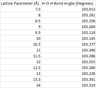
The calculations are done with a cubic unit cell. In order to understand the effects that the images have on each other, we vary the lengths of the cube sides from 7.5 Å to 14 Å in increments of 0.5 Å. Each one of these calculations is done with just the gamma k-point and an energy cutoff of 400 eV.

Fig. 2: Variation of the H-O-H bond angle with respect to the lattice parameter.

By looking at the H-O-H bond angle, we can get an idea of how big the unit cell needs to be in order to minimize the role that the images play in the geometry optimization. Fig. 2 shows that this angle changes rapidly at first, but then starts to stabilize at around 12 Å. A table of the data seen in this plot can be found in the appendix. Unit cells with lattice parameters larger than 12 Å take longer to converge, so this is the lattice parameter that will be chosen for the rest of the calculations.

In order to further confirm that this is an appropriate unit cell size, we can also check to see if the energy has converged.

Fig. 3: Energy of the unoptimized water molecule vs the lattice parameter.

The energy changes by less than 0.1 eV once the lattice parameter is greater than 9 Å. Exact values for these calculations are provided in the appendix.

Energy Cutoff

The calculations for testing the energy cutoff were again done with a k-space sampling that only includes the gamma point. We observe changes in the H-O-H bond angle in increments of 20 eV from 400 eV to 660 eV. A table of these calculations can be found in the appendix.

Fig. 4: Variation of the H-O-H bond angle with respect to the energy cutoff.

As can be seen in fig. 3, the H-O-H bond angle does not have any significant change past an energy cutoff of 600 eV.

We can once again further verify that this is an acceptable energy cutoff by looking at the energy of the unoptimized water molecule as we increase the energy cutoff.

Fig. 5: Energy of the unoptimized water molecule vs the energy cutoff.

The energy changes by less than 0.1 eV between an energy cutoff of 560 eV and 600 eV. Exact values for the energies can be found in the appendix.

K-space Sampling

Due to the high symmetry and size of the unit cell, one should expect that the k-space sampling will have little to no impact on the final geometry of the water molecule centered at the origin in real space. We observe that there is no change in the H-O-H bond angle when using 1, 4, or 14 k-points for energy cutoffs of 400 eV as well as 600 eV.

Testing Initial Conditions

Different initial conditions, i.e the initial bond lengths and the bond angles, provide insight into how the geometry optimization is performed. Given a sufficiently large box size and energy cutoff, one would expect that the water molecule should always converge to the optimal geometry regardless of the initial conditions because it has only one stable configuration. For more complicated molecules, it is important to consider the possibility that there are other stable isomers.

Table 1: Effects of initial conditions on the final geometry of the water molecule and the number of optimization steps. All calculations are run with a lattice parameter of 12 Å and an energy cutoff of 600 eV with only the gamma k-point.

The results in table 1 show that starting with an initial angle of 180° will only cause a change in the two bond lengths. This is because the forces between the atoms only need to be minimized along a line instead of within a plane. Furthermore, we see that starting with two different bond lengths will cause the optimization procedure to take longer to achieve the same result. One can see that achieving what appears to be the most physical result in the shortest amount of steps requires the two initial bond lengths to be the same, while the initial angle must be less than 180°.

Discussion

In order to have accurate geometry optimization calculations of molecules with a plane wave basis set, the chosen unit cell must be sufficiently large so that images of the molecule do not interact. Furthermore, it is important that the cell not be arbitrarily large such that the computation time becomes unreasonable. Once this parameter has been tested, investigations into a suitable energy cutoff can be performed. Convergence can be tested yet again by varying the number of k-points. For a small molecule such as water, just the gamma point was shown to be sufficient. Finally, by utilizing symmetries in the system it is possible to decrease the number of optimization steps. However, one must be careful to make sure that the final results are physical, and to also explore the possibility of other stable isomers.

References

  1.  Hoy, A. R. & Bunker, P. R. A precise solution of the rotation bending Schrödinger equation     for a triatomic molecule with application to the water molecule. Journal of Molecular                 Spectroscopy 74, 1–8 (1979).
  2.  Clark, S. et al. First principles methods using CASTEP. Z. Kristallogr. 220, 567–570 (2005).
  3.  Perdew, J. P; Burke, K; Ernzerhof, M. Phys. Rev. Lett. 1996, 77, 3865-3868.

Appendix

Table A1: H-O-H bond angle as a function of the lattice parameter.

Table A2: H-O-H bond angle as a function of the energy cutoff.

Table A4: Energy of the unoptimized water molecule with respect to the energy cutoff.

Table A3: Energy for the unoptimized water molecule with respect to the lattice parameter.

Determination of the Optimal Geometry of Water Molecules

In this project, the optimal geometry of individual water molecules was found using CASTEP geometry optimization calculations [1]. These calculations utilized the GGA Perdew Burke Ernzerhof (PBE) functional and the Koellig-Harmon atomic solver, which was used to find  psuedopotentials for the hydrogen 1s1 electrons and on the oxygen 1s2 2s2 2p4 electrons. In these calculations, the H atoms had the 1s1 electrons used as valence electrons and the 2s2 2p4 electrons of the O atoms were used as the valence electrons. Additionally, the geometry optimization calculations were performed with a 0.01 eV/Å force tolerance.

Fig. 1. Water Molecule

Determination of Lattice Size

Because CASTEP relies on plane waves, one must construct a lattice to find a molecule’s geometry. Additionally, this lattice must be large enough that the neighboring molecules do not interact in a way that would significantly alter their geometry.  Thus, in performing these calculations, one must ensure that their chosen lattice constant is sufficiently large that the molecule geometry converges past the desired tolerance. In this case, the atomic distances converged to 0.01Å with a 9Å lattice constant, which was used for the primary calculation.

(a)

(b)

Fig. 2. Graph of atomic distances vs. Lattice Constant for (a) OH (b) HH.

Table 1. Effects of lattice constant on bond lengths and angles.

Determination of Energy Cutoff

In order to find the desired energy cutoff for our calculations, the energy cutoff was varied until the distances between atoms converged to 0.01Å. A 435eV cutoff energy was found to be sufficient, and a cutoff of 630eV was used for the main calculation.

(a)

(b)

Fig. 3. Graph of atomic distances vs. cutoff for (a) OH (b) HH.

Table 2. Effects of energy cutoff on atomic distances

Determination of k-space Sampling

To ensure that our k-space sampling was large enough, we checked that the atomic distances converged to 0.01Å at the desired sampling. A sampling of 2x2x2 points, which, after symmetry considerations, resulted in a 4 point total sampling, was found to be sufficient.

(a)

(b)

Fig. 4. Graph of atomic distances vs. cutoff for (a) OH (b) HH.

Table 3. Comparison of total k-space sampling and atomic distances. A single k point was found to be sufficient, and a 4 point (2x2x2) sampling was used in the primary calculation.

Symmetry Considerations

When performing geometry optimization calculations, one must carefully consider the symmetry of the system – in the event that the molecule contains certain symmetries, the forces between atoms may cancel and result in a calculation-breaking local minimum. In this case, when the calculation is performed with the hydrogen atoms perfectly symmetric about an axis (see fig. 5), the calculation fails; the geometry remains symmetric, with OH bond lengths of 0.943Å. However, by starting from slightly modified, symmetry breaking starting positions, we can find values that properly converge.

Fig. 5. A crystal of symmetric H2O molecules with a 9Å lattice spacing. The symmetry about the central oxygen atom causes the calculation to converge to the wrong value.

Conclusion

Utimately, these calculations found that H2O has an OH bond length of 0.97Å with a bond angle of 104.2 degrees. These closely match previously found experimental data  of 0.9572Å and 104.52 degrees [2].

References

Exploring influence of molecular initial guess geometry on optimization

In this project, H2O and HCN molecules were optimized starting with different initial structures, in order to explore the effect of initial structure guess on optimization process and final optimized geometry.

Methods
The geometry optimization was performed in Material Studio with CASTEP Calculation Package [1]. The functional of Perdew Burke, and Ernzerhoff was employed [2]. Since H2O and HCN are two dimensional molecules, two degrees of freedom, including bond length and angle need to be considered for optimization.

Determine Lattice Parameter
To mimic a gas phase molecule, a supercell representing empty space needs to be built. Fig. 1a shows the energy change with lattice parameter and Fig. 1b shows the computational time change with lattice parameter. Considering computational cost and accuracy, lattice parameter of 10 Å was chosen for further study, as energy change between 10 Å and 9 Å is only 0.001 eV, and energy change between 10 Å and 12 Å is also 0.001 eV.


Fig 1 a, Energy change with lattice parameter; 1 b, computational time with lattice parameter

Determine Energy Cutoff
Similar to lattice parameter determination, energy cut-off determination was based on computational cost and accuracy, which were shown in Fig 2. Energy cut-off of 750 eV was chosen for further studies as energy tolerance is less than 0.005 eV.


Fig 2 a, Energy change with energy cut-off; 1 b, computational time with energy cut-off

H2O structure
Influence of initial bond length on optimization
In this section, we will explore how initial O-H bond length affect optimization by fixing H-O-H bond angle 110.37 degree and changing H-O bond length.

Fig 3. Initial structure of H2O molecule
We can see from Table 1, the optimized O-H bond length is 0.97 Å. As initial guess molecule geometry getting closer to the optimized molecular geometry, the computational time and number of iteration will decrease. There is no big difference on final energies and optimized H-O-H bond angle for different initial structures.

Influence of initial bond angle on optimization
In this section, we will explore how initial H-O-H bond angle affect optimization by fixing H-O bond 0.97 Å, as shown in Fig 4.

Fig 4. Fixed H-O bond length and changing H-O-H bond angle
From Table 2, we can see the optimized O-H bond length is 0.97 Å and H-O-H bond angle is 104.23. As initial guess molecule geometry getting closer to the optimized molecular geometry, the computational time and number of iteration will decrease. There is no big difference on final energies and optimized O-H bond length for different initial structures.

HCN structure
Influence of initial H-C bond length on optimization
In this section, we will explore how initial C-H bond length affect optimization by fixing C-N bond length of 1.51 Å and H-C-N bond angle of 180, as shown in Fig 5.

Fig 5. Fixed C-N bond length and H-C-N bond angle
We can see from Table 3, the optimized C-H bond length is 1.075 Å and H-C-N bond angle is about 179.9 degree. As initial guess molecule geometry getting closer to the optimized molecular geometry, the computational time and number of iteration will decrease. There is no big difference on final energies and optimized C-N bond length for different initial structures.

Influence of initial H-C-N bond angle on optimization
In this section, we will explore how initial H-C-N bond angle affect optimization by fixing C-N bond length of 1.159 Å.
It is worth to note that in Table 4, the first 4 rows we were attaching H to N in order to make the corresponding angle. It turned out that such initial structure will generate C-N-H molecule after optimization, as shown in Fig 6. From the first 4 rows, we can conclude as initial geometry getting closer to optimized structure, the optimization time and number of iteration will decrease. But for the last row data, we attached H to C atom and H-C-N angle is very close to optimized structure, we found energy of H-C-N molecule is lower than C-N-H molecule.


Fig 6. optimized C-N-H molecule from Table 5 first 4 rows data.

Conclusion
A good estimate of atomic geometry will greatly increase DFT calculation speed.

Reference
[1] “First principle methods using CASTEP” Zeitschrift fuer Kristallographie 220(5-6) pp. 567-570 (2005)
[2] Perdew, J. P; Burke, K; Ernzerhof, M. Phys. Rev. Lett. 1996, 77, 3865-3868