A month ago I have posted a short piece about developing trichogen cells that resemble spermatozoa. It is always good to see things from different perspectives, so today I post a CLSM media file showing the developing trichogen cells (see Smart Box below) on the third metasomal sternite of the white eyed pupa of Bombus impatiens.
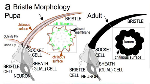
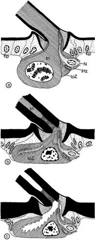
| Box 1. The fate of the trichogen cell The mature bristle (seta) is a hollow structure in Drosophila that corresponds to a single sensory neuron (NEURON) surrounded by a thecogen cell (SHEATH (GLIAL) CELL), a trichogen cell (producer of the hollow bristle, BRISTLE CELL) and a tormogen cell (producer of the socket membrane, a conjunctiva trough which the bristle is flexibly connected to the rest of the cuticle SOCKET CELL).  At the early stage of the development (white eyed pupal stadium), the trichogen cell grows a finger-like projection (by reorganization of actin and microtubule cytoskeleton) and extract cuticle around this projection (chitinous surface).  In later developmental stages (when the cuticle is able to support the developing bristle) the cell projection is redrawn, leaving an empty lumen surrounded by the hardened bristle cuticle (chitinous surface). The general morphology of the mesonotal bristle in the bow fly (Calliphora vicina, Diptera: Calliphoridae; see figure at left) is different from this scheme (Keil 1978). As in Drosophila, the calliphorid mesonotal trichogen cell gives rise a cell projection and starts to develop the surrounding cuticle (Figure 2 a). Unlike in Drosophila, the cell not only redraws the projection, but entirely disappears during later developmental stages (Figures 2b, c). The tormogen cell, on the other hand, enlarges and eventually surrounds the neuron and gives rise the receptor lymph cavities. In Calliphora the tormogen cell has taken over the “spatial” function of the trichogen cell and represents the main cell type in adult bristles. |
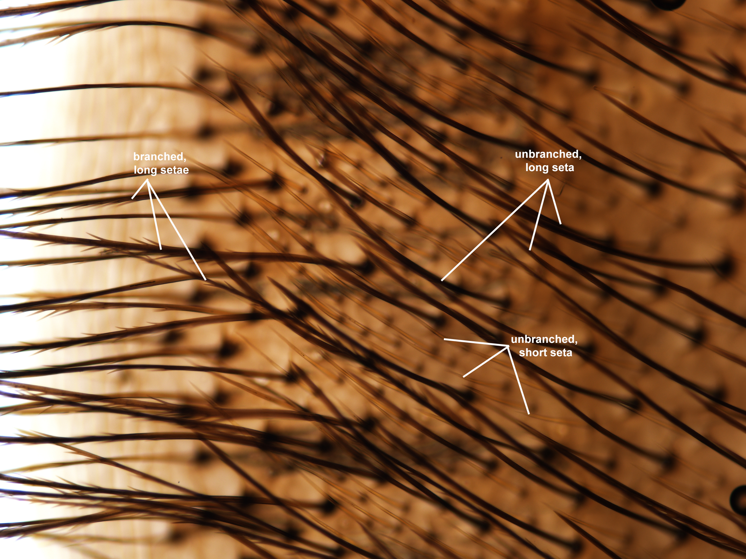
There are two bristle types on the third sternite of the bumble bee: Long and brownish (melanized) bristle (branched, long seta; unbranched, long seta: Fig. 3) corresponding to larger trichogen cells (trichogen long: Fig. 1.) and shorter, transparent (not melanized) bristle (unbranched, short seta: Fig. 3), corresponding to smaller trichogens (trichogen short: Fig. 1). Beside the seta-producing trichogen cells, numerous other, smaller epidermal cells are visible on Fig. 1.

The long and more melanized setae are branched in the posterior half and unbranched in the anterior half of the hairy part of the sternite.

We have gathered another CLSM image that reveals that the long, melanized setae in adults correspond to one large and a few smaller epidermal cells.

Normally I would assume that the large cell is the trichogen cell, while one of the smaller cells (perhaps the drop-like one that is connected to the setal base) is the tormogen cell, but after reading Keil 1978 (and knowing that cell fate is different even within Diptera), I would not do this generalization without a thorough examination of the developing bumble bee integument.
Leave a Reply