This post is the fourth in a short blog series called “Know your Insect”. The images and descriptions are written by Entomology graduate students enrolled in a seminar of the same name.
By: Kyle Burks
The insect circulatory system is very different from that of mammals. Insects do not have lungs and they do not circulate oxygen through blood. Instead, they have a series of openings along theirs sides called spiracles. The spiracles open up into a system of air-filled tubes, the tracheae. Tracheae terminate in very fine branches called tracheoles which supply oxygen directly to every cell.
Insects can have up to two spiracles on the thorax and eight on their abdomen. Thoracic spiracles have a different morphology from abdominal spiracles. Abdominal spiracles can have up to two muscles that open and close them (dilator and occlusor; Fig. 1), though thoracic spiracles usually have a single closer (occlusor) muscle (Figs 2-5). Muscles are not necessary for opening spiracles, though. When relaxed, the spiracles are at least partially open (Chapman 2012). This prevents suffocation if consciousness is lost.

Thoracic spiracles have diverse morphology (Hassan 1944). One of the more common systems, at least among Hymenoptera, is a single muscle which tugs on a flexible ring at the beginning of the trachea. We took images of the first thoracic spiracle of several undescribed species of Nearctic Dendrocerus (Hymenoptera: Megaspilidae) using Confocal Laser Scanning Microscopy (CLSM). CLSM excites the specimen with a laser and captures the fluorescence that results.
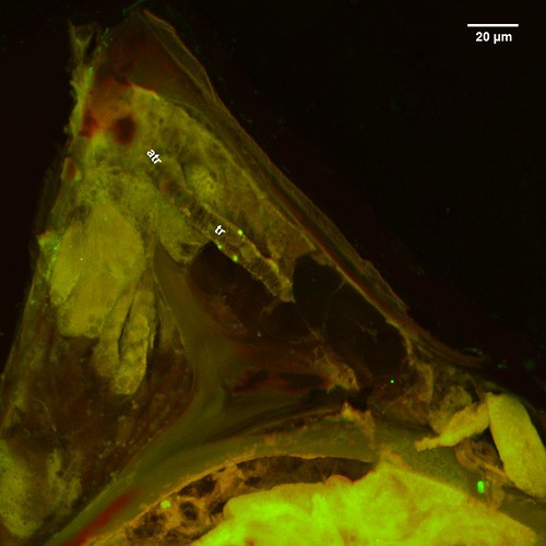

CLSM splits the fluorescence into multiple channels based on their wavelength. Since different kinds of structures auto-fluoresce in different wavelengths, the color in the CLSM image encodes useful information. Soft structures like muscles, fat bodies, glands, and conjunctiva appear green, while harder structures such as the cuticle or chitinous structures appear red with 488 nm excitation laser. Areas with an abundance of the elastic protein resilin fluoresce the most strongly with the 405 nm laser in the blue range.
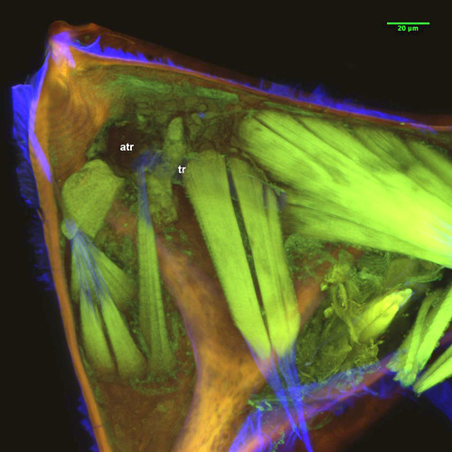
Between the opening of the spiracle and the start of the trachea, there can be up to two chambers with a hardened, thickened wall called atria (singular: atrium; Tonapi 1957). Atria are common among insects. In the CLSM images of Dendrocerus, we can see that a muscle inserts between the atrium (atr: Figs 2, 4) and the trachea (tr: Figs 2, 4). The bright blue in this region is the flexible tendon of the occlusor muscle and the flexible ring that closes the trachea.
The group of wasps that I study, Dendrocerus, sometimes have enormous atria. Why might they have such large atria though? What functions might a large atria such as this be capable of? Dendrocerus are extremely small wasps, and as such, are capable of being trapped in water droplets. This is also a danger experienced by some thrips, which have a modification to the outside of their spiracles which trap air bubbles around the the spiracle to prevent accidental drowning (Wiesenborn 2014). Could this explain the large atria of Dendrocerus? Might it act as an emergency air reservoir? Perhaps it may help maintain humidity instead. By having a large, round forechamber the tracheal system does not open up directly to the outside, which could help prevent water loss.
Do you have any ideas on what the function of these large atria could be? Leave a comment!
Leave a Reply