This post is the sixth in a short blog series called “Know your Insect”. The images and descriptions are written by Entomology graduate students enrolled in a seminar of the same name.
By: Luca Franzini
Insects live in a world dominated by scent. Chemicals play a fundamental role in most aspects of their complex, fascinating lives and are frequently actively produced by the insects themselves. For example, a queen honey bee rules over tens of thousands workers just by releasing a single, specific compound, the queen mandibular pheromone 9-oxo-(2E)-decenoic acid, which suppresses the ovarian development of her subjects (Hoover et al., 2003). Furthermore, females of several species of moth will attract males over long distances by emitting species-specific sex pheromones. Only males from their species will come to court them. However, what do all these chemicals, eliciting such complex behaviours, have in common? Well, they are all produced by glands.

Glands in insects are found spread across their whole body, in their mandibles, in their brain, in their gut. Their role is as varied as insects themselves, and range from mate attraction to defense to complex social communication. While most glands are found across all insect orders, several taxa will often possess their own unique glands, which will also vary in their uses and contents across the various species.
The Dufour’s gland is found only in Hymenoptera, in the suborder Apocrita. It was first described by Léon Jean Marie Dufour in 1841 and for a long time it was known as the alkaline gland. This name derived from the misconception that, together with the acid gland (now known as the venom gland), it was associated with stinging in bees and wasps. It was thought that its secretions activated the venom, by mixing with those from the acid gland, or that they would neutralize the acidic ones left inside the sting after a bee or wasp had stung an enemy.

While it is true that the Dufour’s gland is associated with the sting apparatus in Aculeata, it has no role in stinging or venom production and its duct opens into the dorsal vaginal wall (Billen, 1987). The only exception to this is found in the family Formicidae, where the gland opens into the sting bulb, but still with no involvement with the main function of the sting itself (Billen, 1987). It develops as an invagination of the dorsal vaginal wall (membrane connecting the ventral margins of the dorsal valves) and like the other accessory glands of the sting apparatus, the venom gland and the spermatheca, it is of ectodermal origin. Shaped like a long, slender tube, it displays conspicuous folding of its epithelium, which gives the gland an accordion-like appearance (Billen, 2006). The gland cells are lined with cuticle, as in other class I epidermal glands, through which their secretions must go through to reach the lumen.

The gland content varies across species, but in most taxa it is mainly composed by long-chain saturated and unsaturated hydrocarbons, a condition that appears to be the ancestral one for the Dufour’s gland (Mitra, 2013). These compounds are not synthesised by the gland itself, but are produced by specialised secretory cells called oenocytes (Makki et al., 2014).
These cells are found in clusters in the abdomen and sometimes in the thorax of most Pterygota and are often in association with fat bodies. They possess a well-developed smooth endoplasmic reticulum and play a key role in lipid metabolism, detoxification, and synthesis of hydrocarbons and other lipid-derived substances (Makki et al., 2014). From the oenocytes, these compounds are released into the haemolymph from which they reach the Dufour’s gland and the cuticle where they create a waxy layer. This coating on the cuticle is usually a mixture of hydrocarbons, sterols, fatty acids, fatty alcohols, and wax esters. The function of this coating is primarily as a barrier against desiccation. Moreover, the profile created by these chemicals is usually species-specific and it is involved in chemical communication (Mitra, 2013). Interestingly, the content of the Dufour’s gland has been found to match closely the composition of this cuticular waxy layer in a variety of species, including bumble bees and the social wasp Ropalidia marginata (Oldham et al., 1994).
The cuticular hydrocarbon profile of social Hymenoptera is of fundamental importance for in-nest dynamics, as it represents one of the main components of their complex chemical communication system. Not only does the profile tend to be species-specific, but it is often also colony specific. This provides a barrier against potential intruders. Nonetheless, communication is just one of the many uses for the Dufour’s gland, some of which can be quite peculiar. For example, in parasitic wasps of the superfamily Ichneumonoidea, female wasps will use secretions from this gland to mark their hosts after laying their eggs in it to avoid competition from related individuals (Mitra, 2013).

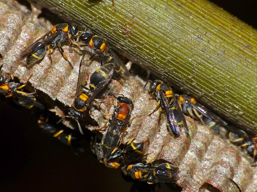
In social wasp from the subfamily Stenogastrinae, the gland secretes a white, gooey mass, called abdominal substance, which serves a variety of functions (Turillazzi, 1991).
It is placed around the pedicel of their nests where it acts as an ant repellent, but it is also used for liquid food storage and, most importantly, as the base onto which the eggs are laid.
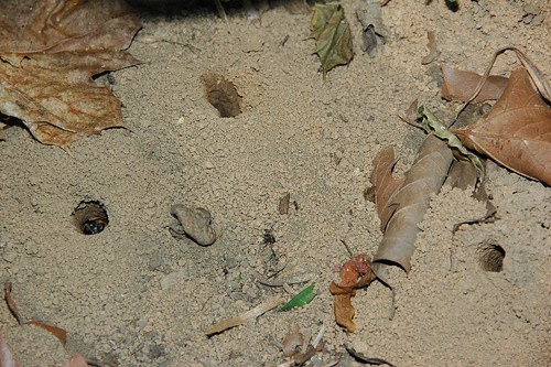
In solitary bees, in particular in ground-nesting species (e.g., Colletidae, Andrenidae, Halictidae), the gland became enlarged during the construction of the brood cells, occupying large part of the metasoma (Lello, 1976). This is due to the fact that the Dufour’s gland in these species secretes macrolytic lactones, a form of natural polyesters, which are used in brood cells lining (Hefetz, 1987). This cellophane-like coating prevents desiccation, flooding of the cells, and it has antimicrobial properties.
Hypertrophism of the Dufour’s gland is also found in dulotic ants (D’Ettorre et al., 2000). Also known as slave-making ants, they raid other species’ nests, capture their brood and after bringing it back to their nest they enslave the emerging workers. To help in their raids, they rely on the propaganda substances produced by the Dufour’s gland, which mimic the alarm pheromone of the host and will cause alarm among the defending workers, generating panic and allowing the raiders to pillage the nest (Buschinger, 2009).
Hacking into another species’ communication system is more common than one would think, and it is rarely done without ill-intent. For instance, males of several species of the kleptoparasitic genus Nomada will, while mating, coat the female with secretions from their mandibular glands (Tengö and Bergström, 1977). These secretions mimic those of the Dufour’s gland of the host species and help the female avoid confrontation with the host while entering its nest (Mitra, 2013).
Another well-known example of this kind of hacking is social parasitism, a fascinating lifestyle where a species will exploit the social structure of another for its own benefit, typically to rear its brood.
Social parasites are common within the various groups of social Hymenoptera, and bumble bees are no exception. There are about 250 species of bumble bees (Apidae,Bombus), about 30 of which are socially parasitic. With the exception of two species, all the remaining parasitic ones can be found grouped in the subgenus Psithyrus. Members of this subgenus, commonly referred to as cuckoo bumblebees, are morphologically and physiologically adapted to a parasitic lifestyle and rely on various chemical strategies to infiltrate their host colonies and usurp them (Fisher and Sampson, 1992).

Chemical mimicry is perhaps the most fascinating among the strategies used by these parasites. It has been demonstrated that some species of cuckoo bumblebees actively mimic the cuticular hydrocarbon profile and Dufour’s gland content of their host species (Martin et al., 2010). This allows the usurper to take over the role of dominant reproductive female, the queen, with relative ease and maintain it long enough for its offspring to fully develop inside the host colony. When this mimicry is not possible or achieved, as in the case of generalists parasitising more than one host species, some species will sneakily rely on worker repellents, like dodecyl acetate, secreted from their Dufour’s gland (Zimma et al., 2003; Martin et al., 2010).
My research aims at further investigating the chemical strategies employed by cuckoo bumblebees during their rule of a host colony. I will look into various aspects of the usurpation process, from chemical mimicry to the use of worker repellents, in a wider range of species and I will investigate how the different developmental stages and sexes of the social parasite cope with life inside another species’ nest. Even though these are just few of the questions left unanswered about the cuckoo bumblebees’ life history, and only a small portion of those I would like to answer with my future research, I hope my work will shed some light on the still poorly-understood life of these amazing insects.
Leave a Reply