Since we published our first Confocal Laser Scanning Microscopy (CLSM) image/media file in 2009 we often apply this imaging technique to visualize minute and intricate anatomical structures of insects.
Sometimes, however, we would like to image larger specimens that do not fit into the picture of the lowest magnification objective (10x). We first faced this problem when conducting a study on the membracid “pronotal wing” (well, pronotum). For this project we tried to stitch individual images together with, let’s say, moderate success (you certainly can see the borders of individual images).

Later we made another trial on a head louse with similar results.

I am sure we could have improved the quality of panorama imaging with the ordinary CLSM, but we didn’t really need this option … we were (and mostly are) working with moderately sized insects in the last few years.
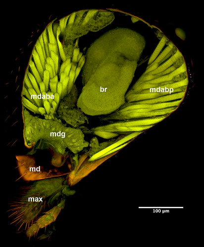
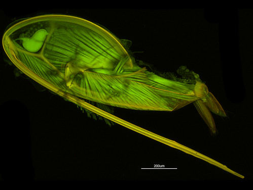
Then we started our project on the ichneumonoid ovipositor. While the ovipositor of even the largest ceraphronoid fit perfectly in the view of a 10x objective (Figure 1) we can hardly squeeze in the basal articulation of an ichneumonoid ovipositor.

Panorama stitching is a well known method in normal brightfield imaging and using the gigapan website we are now able to publish high resolution images for supporting scientific publications.

But how could we produce good quality, high resolution CLSM micrographs? Would it be once possible to publish a single high resolution CLSM image instead of a low resolution overview and a higher resolution “detail” image?

As usual, the Microscopy and Cytometry Facility of the Huck Institute of the Life Sciences provides a solution: the Olympus FV10i.

With this small “R2D2” we can finally make really high quality CLSM panorama images.