
Ultrasound Twinkling of Kidney Stones
The color Doppler ultrasound twinkling artifact, which highlights hard objects such as kidney stones with rapidly changing color, has the potential to improve kidney stone detection; however, its inconsistent appearance has limited its clinical utility. Recently, it was proposed that stable bubbles trapped in the crevices on the kidney stone surface cause twinkling because hyperbaric pressures eliminated twinkling on ex vivo kidney stones. During her postdoc, Dr. Simon showed that these bubbles may be internal as well as on the kidney stone surface and that respiratory gas composition alters the presence and amplitude of kidney stone twinkling.

BASiL is working with the Urology department at Penn State Hershey Medical Center to investigate whether breathing elevated oxygen enhances twinkling in patients. As the current gold standard for kidney stone detection in the United States is x-ray computed tomography, enhancing the twinkling artifact for kidney stone detection will reduce patient exposure to ionizing radiation.
_______________________________________________________________________
Focused Ultrasound for Tendon Injuries
Traditionally, ultrasound has been used at very low intensities to produce mild heating and elevated blood perfusion to aid in tendon healing. Therapies such as dry needling and shock wave therapy are often used to enhance tendon healing with mixed success. BASiL proposes using the bubbles induced by focused ultrasound histotripsy to create targeted microdamage to the tendon and promote a healing response. The primary advantages of histotripsy therapy over other modalities are that it is completely noninvasive, can be precisely guided with ultrasound imaging, and may improve collagen alignment in the healing fibers. BASiL is collaborating with Dr. Meghan Vidt’s Movement of the Upper Limb and Shoulder Lab (MUSL) at Penn State to develop mechanical models to validate and scale the therapy to large animal models before working in humans.

_______________________________________________________________________
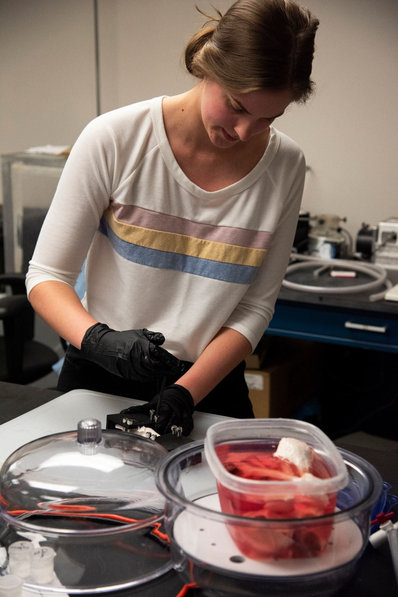
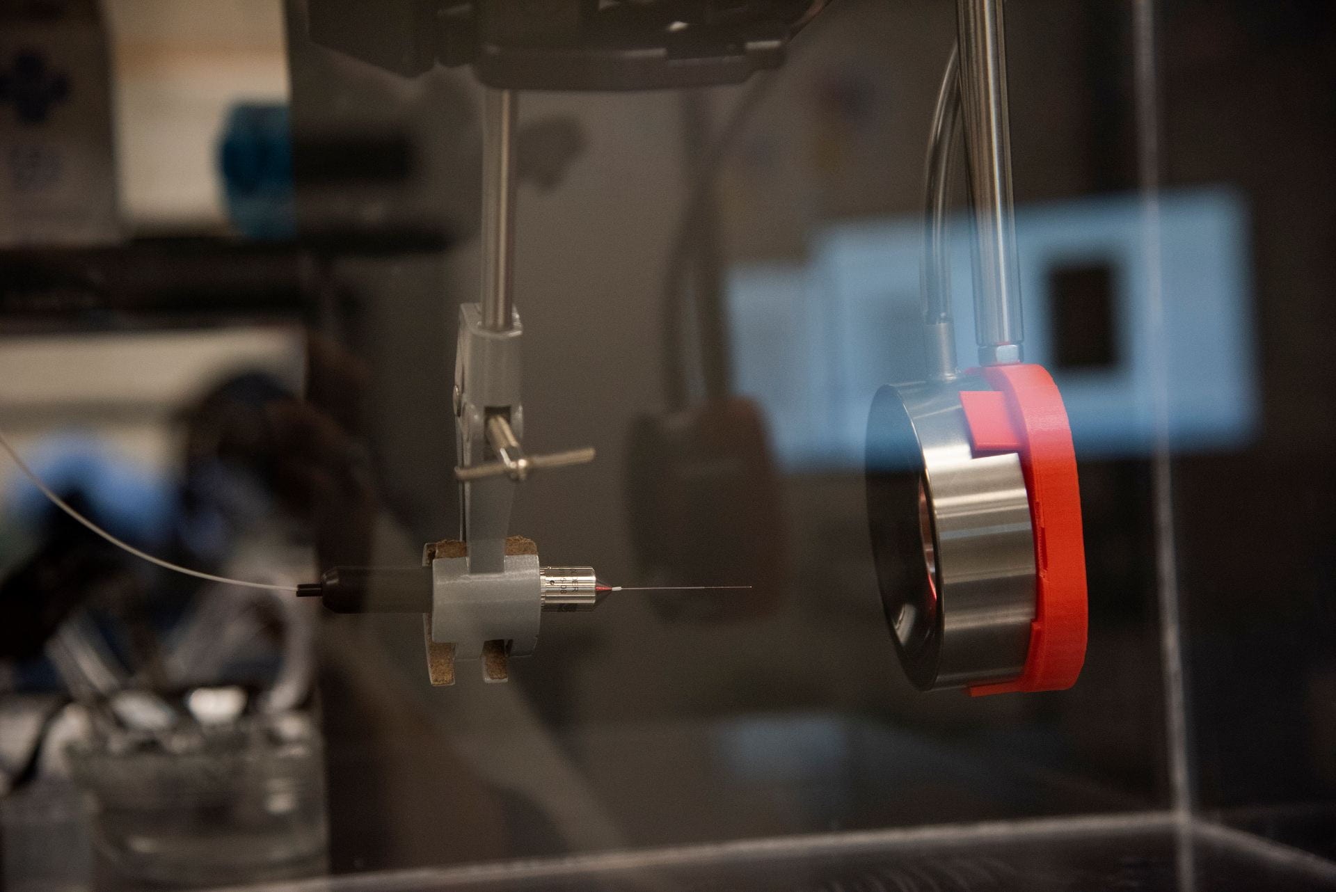
Cavitation-Based Treatment Monitoring
BASiL is currently working on a project to show histotripsy as a potential healing modality for soft tissue, namely tendons. Focused ultrasound at high intensities cause the formation of bubbles within the targeted tissue, which, upon their collapse, creates microdamage and releases healing factors within the area. Quantifying the strength at which these bubbles collapse can be done using passive cavitation detection, where a “listening” transducer receives emission signals from the bubbles. This data can then be post-processed to receive an image indicating the relative strength of the collapse at and near the source. BASiL is working to link the cavitation signal to the desired tissue effect using both passive cavitation imaging and Doppler ultrasound to monitor tissue motion.

_______________________________________________________________________
Ultrasound for the Diagnosis and Treatment of Pathological Biomineralization
Commonly used to aid kidney stone diagnosis, the color Doppler ultrasound twinkling artifact has also been seen in gallstones and some atherosclerotic plaques, suggesting that crystals, regardless of chemical composition, may harbor the stable microbubbles that are hypothesized to cause twinkling. BASiL is currently investigating uric acid, cholesterol, calcium phosphate, and calcium oxalate crystals to determine how crystal chemical composition influences the twinkling artifact. Goals of this project are to improve early diagnosis and explore histotripsy fractionation on crystals as a novel treatment for pathological biomineralization – such as heterotopic ossification.

_______________________________________________________________________
Ultrasound-Guided, Focused-Ultrasound-Mediated Drug Delivery
Several labs at Penn State are already working with BASiL about using ultrasound to image and release drugs or molecules from specific drug delivery vehicles or hydrogels. We are open to additional collaborations if you would like experts in acoustics to consult or work to release drugs from different carriers.