This post is the third in a short blog series called “Know your Insect”. The images and descriptions are written by Entomology graduate students enrolled in a seminar of the same name.
By: Carolyn Trietsch
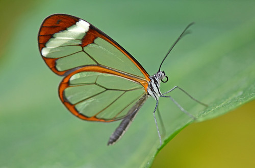
Transparent Cuticle in Insects
The cuticle (the external, acellular layer of the integument) is an integral part of an insect’s anatomy. Not only does the cuticle act as a skin to protect the insect’s internal anatomy from the outside environment, but also as a skeleton to provide support and allow for movement (Rockstein 1974).

Some parts of the cuticle are transparent, and this transparent cuticle can serve many different functions in insects. Transparent cuticle can be found over the eyes and ocelli, allowing light to shine in so that the insect can see. Transparent cuticle can also be found over bioluminescent organs, allowing light to shine out. In fireflies, the transparent cuticle present over the light organ contains nanostructures that actually increase the transmission of light. These cuticular nanostructures have inspired the development of more efficient LED lenses (Kim et al 2012).
Transparent Cuticle and Extra-ocular Photoreceptors
Extra-ocular or extra-retinal photoreceptors are photoreceptors found outside of the eyes and ocelli (Arikawa 1999). Extra-ocular photoreceptors have been found in the telsons of scorpions (Zwicky 1968; Rao 1973) and horseshoe crabs (Hanna et al 1988; Renninger et al. 1997), as well as in the brains of beetles (Fleissner & Fleissner 1992; Fleissner et al. 1993a) and possibly cockroaches (Fleissner et al. 2001). Photoreceptors have even been found in the genitalia of butterflies, playing an important role in ensuring that the male and female are properly aligned during copulation (Miyako et al. 1993; Arikawa & Miyako-Shimazaki 1996; Arikawa 1999).
Transparent cuticle can occur over extra-ocular photoreceptors, allowing light to shine through the cuticle. Pupae of the oak silkworm, Antheraea perny, have a transparent patch of cuticle directly over the brain. When light shines through the patch, it is detected by extra-ocular photoreceptors in the brain that are important for regulating diapause. Based on the photoperiod (how many hours of light and dark), light shining through the transparent patch will stimulate the brain to secrete a hormone that is important for development of the pupa into an adult. In this way, photoperiod is important for regulating the endocrine activity of the brain (Williams & Adkisson 1964).
Semitransparent patches in the abdomens of Conostigmus sp.
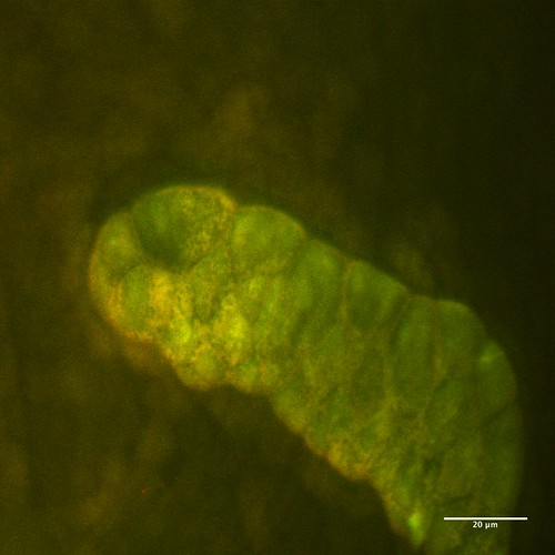
On the abdomens of ceraphronoid wasps, there are two semitransparent patches on the dorsal surface and two semitransparent patches on the ventral surface of the abdomen (Mikó & Deans 2009). The structure and function of these patches are completely unknown. It is possible that these wasps might be bioluminescent, and that there are light-emitting organs underneath the patches. Unfortunately, this cannot be determined from ethanol-preserved specimens; living specimens are required.

It is also possible that there may be photoreceptors underneath the patches. Extra-ocular photoreceptors could be used for orientation during flight, or possibly to help orient towards light sources while burrowing into substrate. The semitransparent patches themselves are concave and have patterns resembling stained-glass windows; it is possible that the patches may have nanostructures that specifically help to focus light on extra-ocular photoreceptors.

In dissecting a Megaspilus sp., we found what could be gland cells underneath the patches. Located next to the semitransparent patches in Ceraphronoid wasps are setiferous patches, characterized by setae. In looking at cross sections of the abdomen, we found what looked like pore canals in the cuticle underneath the setiferous patches. It is possible that light-stimulated glands underneath the semitransparent patches could secrete pheromones onto the setiferous patches. This would be similar to the function of felt patches in wasps of the superfamily Platygastroidea (Mikó & Deans 2009).

We also found an interesting synapomorphy in Ceraphronoidea; whereas other Hymenoptera have multiple muscles that control the telescopic motion of the abdominal segments, in Ceraphronoidea there is only one long muscle that extends throughout the abdomen. This muscle originates at the semitransparent patch- could it be that the muscle requires light or the warmth of sunlight in order to function properly?
This research is exciting in that it is full of potential and can be continued in many different ways. One future step would be to use tip enhanced Raman spectroscopy to analyze the surface of the semitransparent patches. This technique is advantageous because it can be used to detect organic compounds on surfaces without destroying them (Araujo et al 2014).
Another future step to take in this research would be to determine if there are actually photoreceptors correlated with semitransparent patches in these wasps. One way to do this would be to design antibodies specific for opsins, proteins that are associated with photoreceptors, in order to indicate possible locations of photoreceptors within the insect’s body (Lampel et al 2005).

Leave a Reply